A chest CT scan is an imaging examination performed using modern CT scanners that enable accurate visualisation of the organs located in the mediastinum – the area between the head and the abdominal cavity. Computed tomography of the chest cavity makes it possible to diagnose various diseases and injuries, and can also be used to monitor the effects of treatment.
What can a chest CT scan detect?
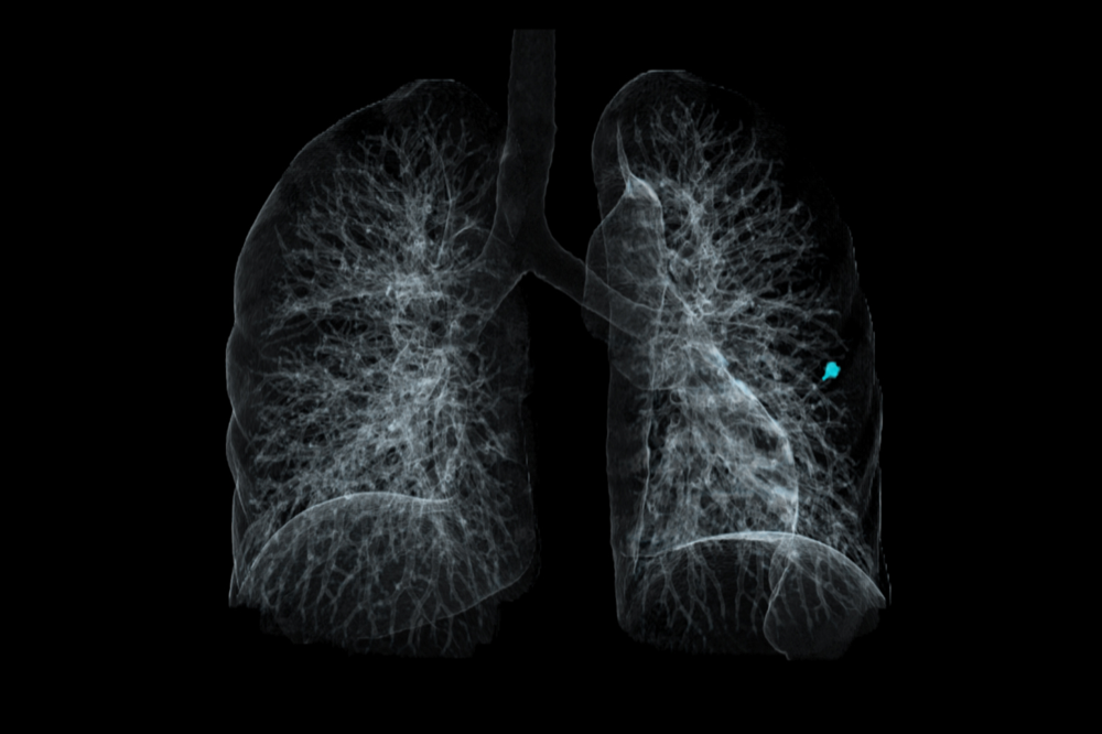
Chest tomography allows an in-depth analysis of the organs inside the chest and its internal structures. It is primarily used to diagnose diseases of the lungs, bronchi, trachea and heart. A chest computed tomography scan can detect the following conditions and perform the following assessments:
- lung tumours, both benign and malignant
- infiltrates in the pericardium, heart or esophagus
- vascular diseases, including aortic aneurysms
- lung diseases such as pneumonia, emphysema, pulmonary fibrosis, fibroid tuberculosis or pneumoconiosis
- lesions following chest trauma, adhesions, damage to the pulmonary parenchyma
- inflammation in the lungs
- pericarditis
- pleural effusion
- lesions in mediastinal lymph nodes
- diseases of the heart and large vessels, including heart defects
- assessment of the patient’s condition after thoracic surgery
- congenital malformations of the trachea, lungs and bronchi
- complications after COVID-19
- vascular diseases
Chest CT scans make it possible to diagnose various conditions and determine their severity, and subsequently monitor the patient’s progress after treatment has begun.
Indications for a chest CT scan
A CT scan of the chest requires a referral from a doctor because it is a radiological examination (one which involves exposure to X-rays). While modern devices such as UIH CT scanners enable radiation doses to be significantly reduced while maintaining high image quality, caution should be exercised with this type of examination.
Indications for a chest computed tomography scan include:
- Diagnostics of pulmonary and bronchial diseases, for instance pneumonia, asthma, pneumoconiosis, lung tumours.
- Evaluation of heart and large vessel disorders including heart defects, aortic aneurysms.
- Examination of lesions after chest trauma.
- Detection and monitoring of cancerous lesions, including assessment of response to treatment.
- Diagnostics of infectious and inflammatory diseases.
- Assessment of the patient’s condition after thoracic surgery.
What does a chest CT scan look like?
A chest CT scan is similar to other examinations performed using CT scanners. The examination itself takes about 15 to 30 minutes. In the case of a chest CT scan with contrast, a contrast agent is first administered, which is infused intravenously via a peripheral venous catheter. The patient must then lie down on a special table, which is slid into the CT scanner bore.
During the scan, the patient usually has to hold his or her breath. Instructions are communicated by the radiologist via intercom – the patient remains in contact with the operators throughout the examination, which enables him or her to immediately signal any problems such as claustrophobia or a possible allergic reaction to the contrast agent.
After a contrast-enhanced chest CT scan, it is advisable to stay at the medical facility for at least half an hour in case an allergic reaction develops. Chest CT results are usually available after a few days because they must be annotated, and this must be done by a radiologist.
Specialised chest CT scans
A chest CT scan can provide a non-invasive alternative to tests such as coronary angiography. High-resolution CT of the chest also enables extremely accurate diagnosis of lung diseases. In this regard, there are two primary specialist CT examinations:
- Pulmonary perfusion CT – a CT scan showing the flow of blood through the lungs.
- Angio-CT of the pulmonary arteries – an examination that can detect pulmonary embolism in the pulmonary artery or its branches.
CT pulmonary angiography (CTPA) is always performed with contrast.
Preparation for a chest CT scan
No special preparation is required for non-contrast-enhanced chest CT scans. The patient should wear loose clothing without metal parts, because these can interfere with the image. For contrast-enhanced chest CT scans, the patient should remain fasting for at least 4 to 6 hours before the examination. He or she, however, should drink plenty of non-carbonated water.
Before administering contrast certain medications must be discontinued, such as metformin, which is used by diabetes patients. Individuals with a history of allergic reactions to the contrast agent should take glucocorticosteroids or antihistamines before the examination. Such patients should inform medical staff of their previous allergic reactions before contrast is administered.
It is also important to note that pregnancy is a contraindication for a chest CT scan, as X-rays can be harmful to the foetus.