The panoramic high-definition perspective empowers accurate diagnosis
The general medical image processing platform covers full-modal, multi-dimensional, advanced clinical applications, helps provide a panoramic high-definition perspective for the analysis of medical images, and empowers accurate clinical diagnosis.
More than 60 advanced applications in multimodality workstation (CT, MR, PET-CT, PET-MR). Helping doctors make accurate and efficient diagnosis in different clinical scenarios such as cardiovascular, oncology, neurology.
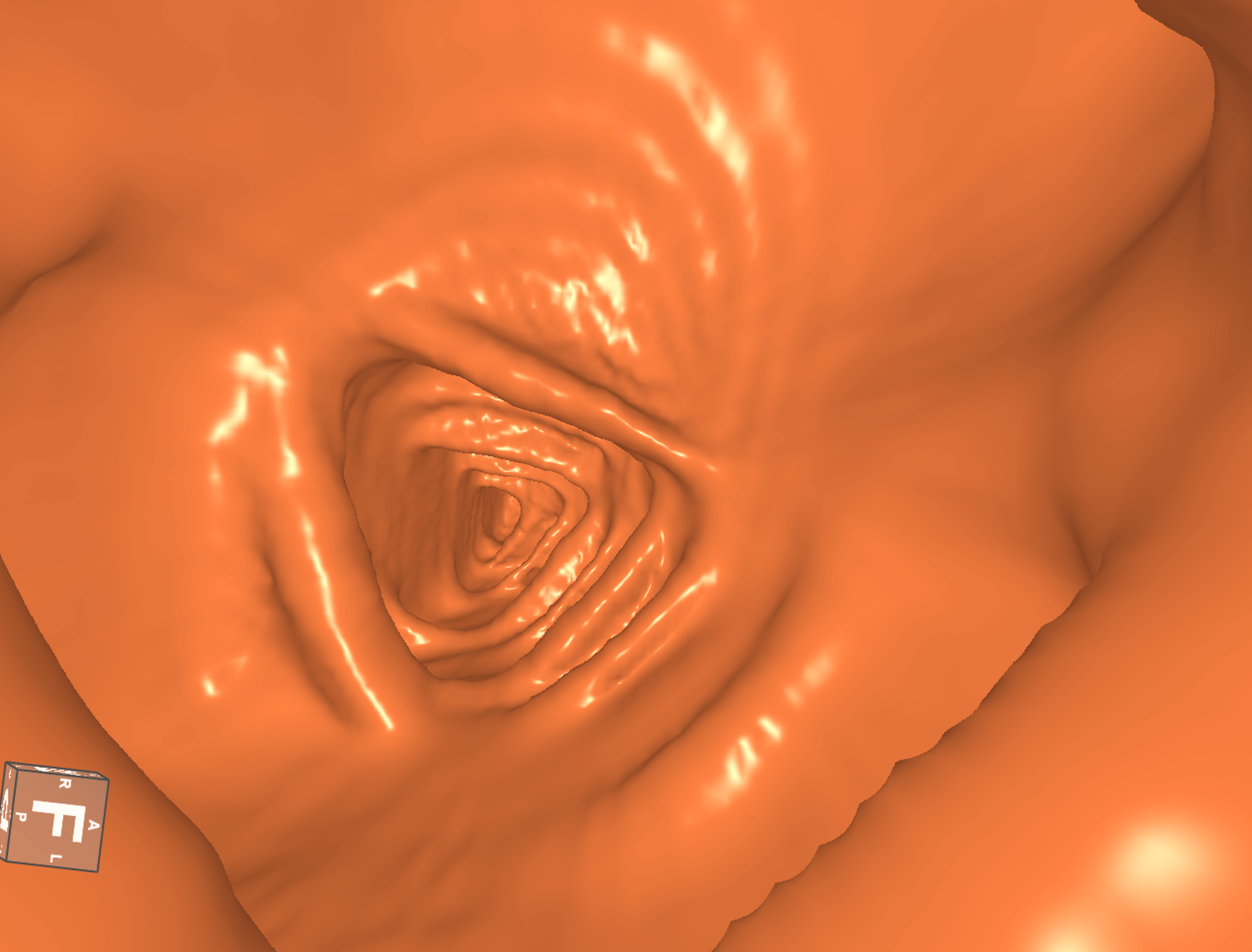
Provide virtual endoscopy view to observe the internal structure of the tissue.
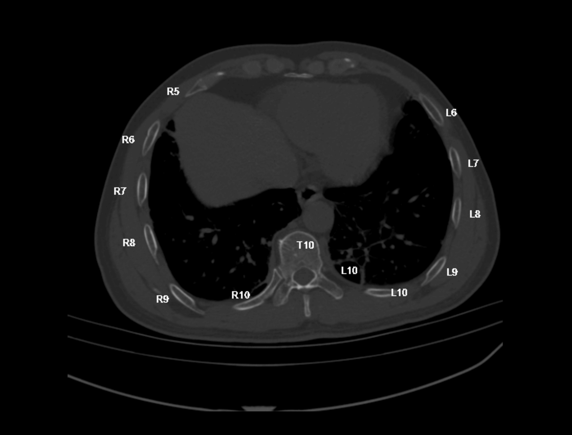
Rapid rib segmentation and extraction are offered to assist in rapid fracture positioning.

Different types of images can be fused and displayed to provide more reference information.
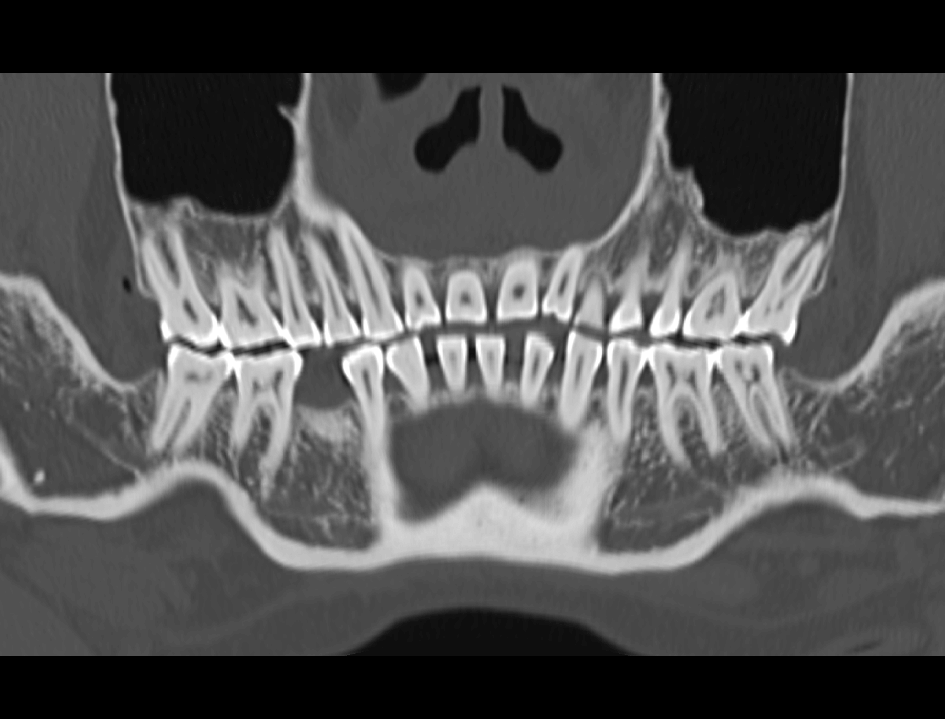
Provide panoramic and cross-sectional views, provide auxiliary reference information for orthodontic treatment plans.
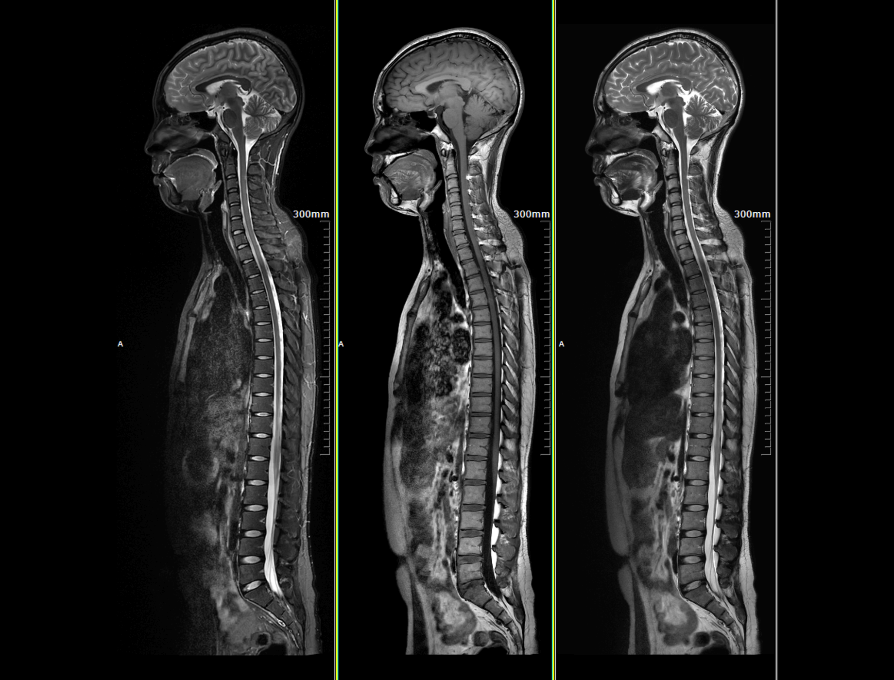
Stitching can stitch several images scanned by segments for panoramic view. Automatic stitching can rapidly gain spine, vessel and whole body stitched images.
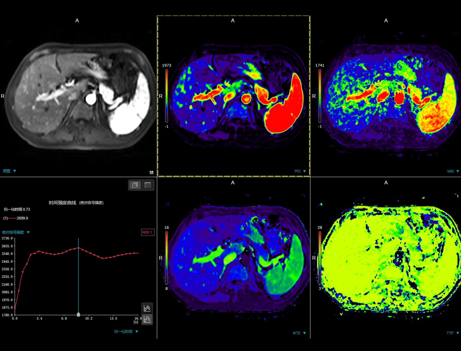
Dynamic Evaluation is used to analyze dynamic magnetic resonance images. It provides mean time intensity curve of ROI and parameter maps of positive analysis or negative analysis.
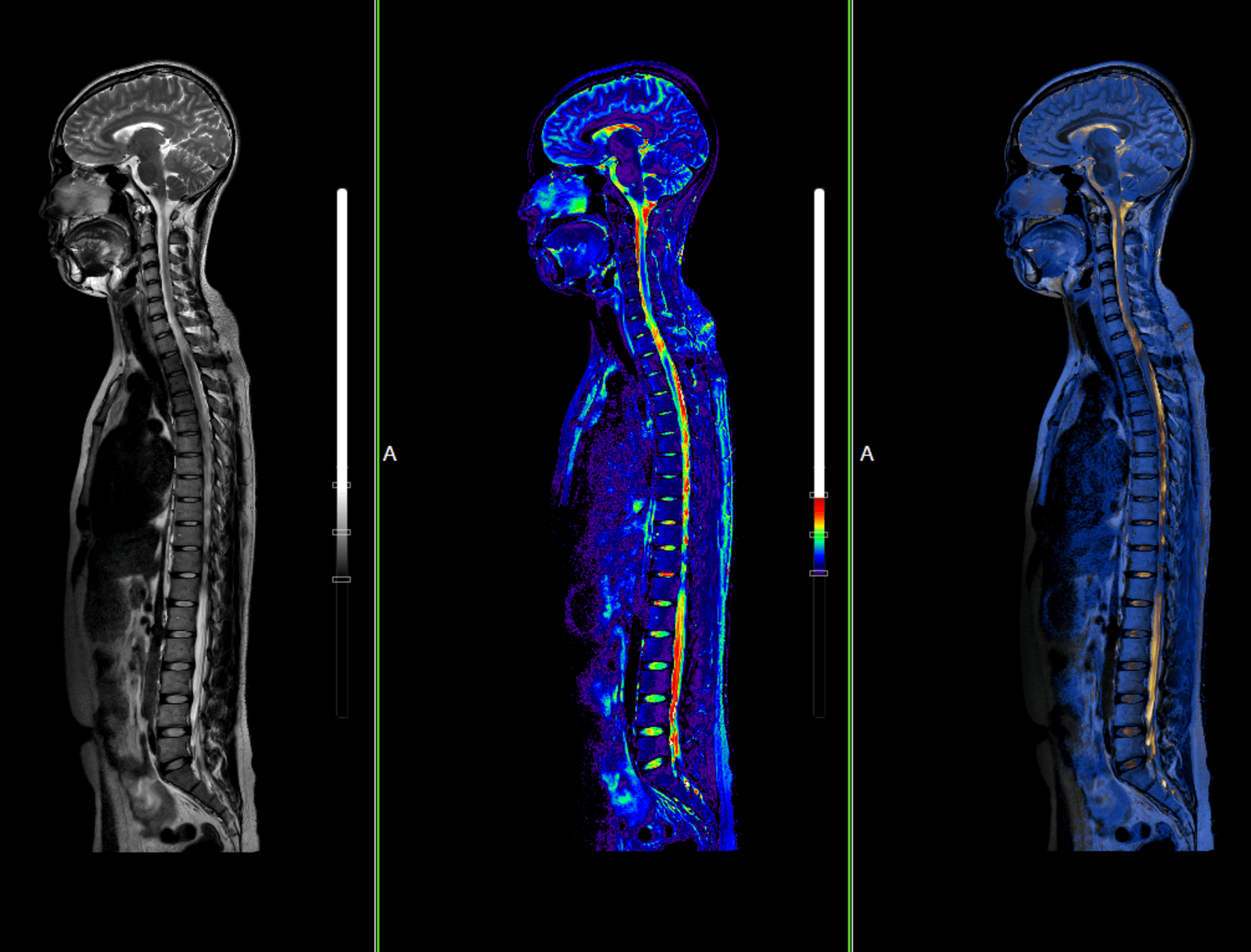
MR image fusion can fuse different images by image registration technology, support to adjust blend ratio,pseudo-color scenarios and reference data.
Inner View offers segmentation algorithms to automatically or manually extract the dividing lines of vessel, and simulates an endoscopy display within the organ by using a 3D display from different angles.

The application provides comprehensive tools for calculating and plotting metabolism activity in a user determined region of interest (ROI) over time and location.
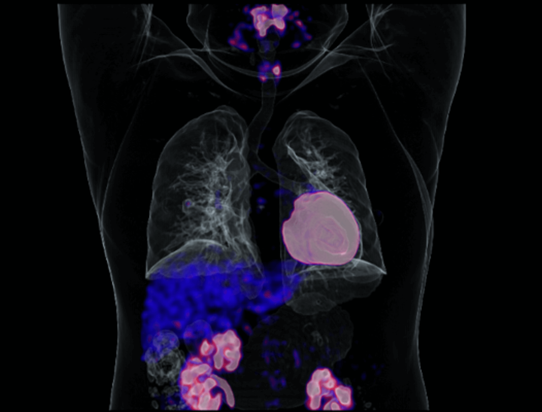
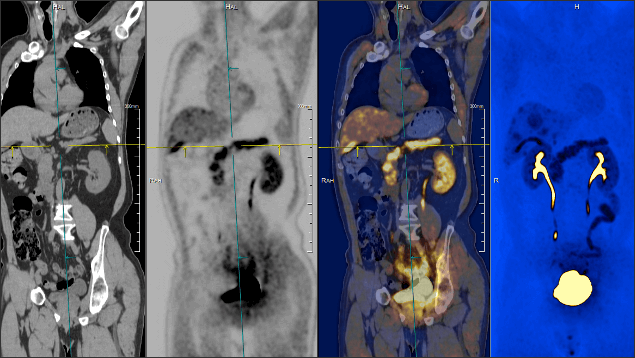
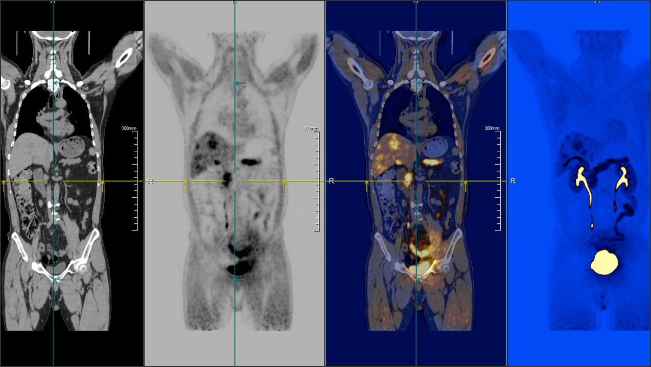
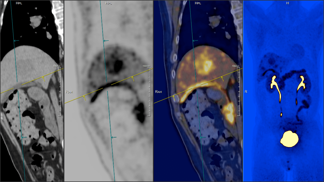
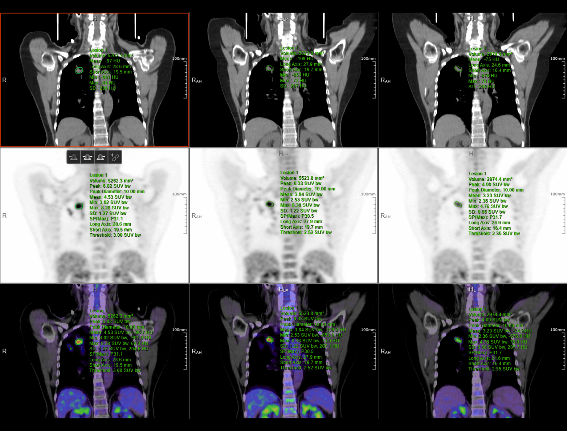
Image Fusion offers quick and configurable protocols for fused PET/CT and PET/MR image reading and convenient common tools to help you achieve the outcomes you need.
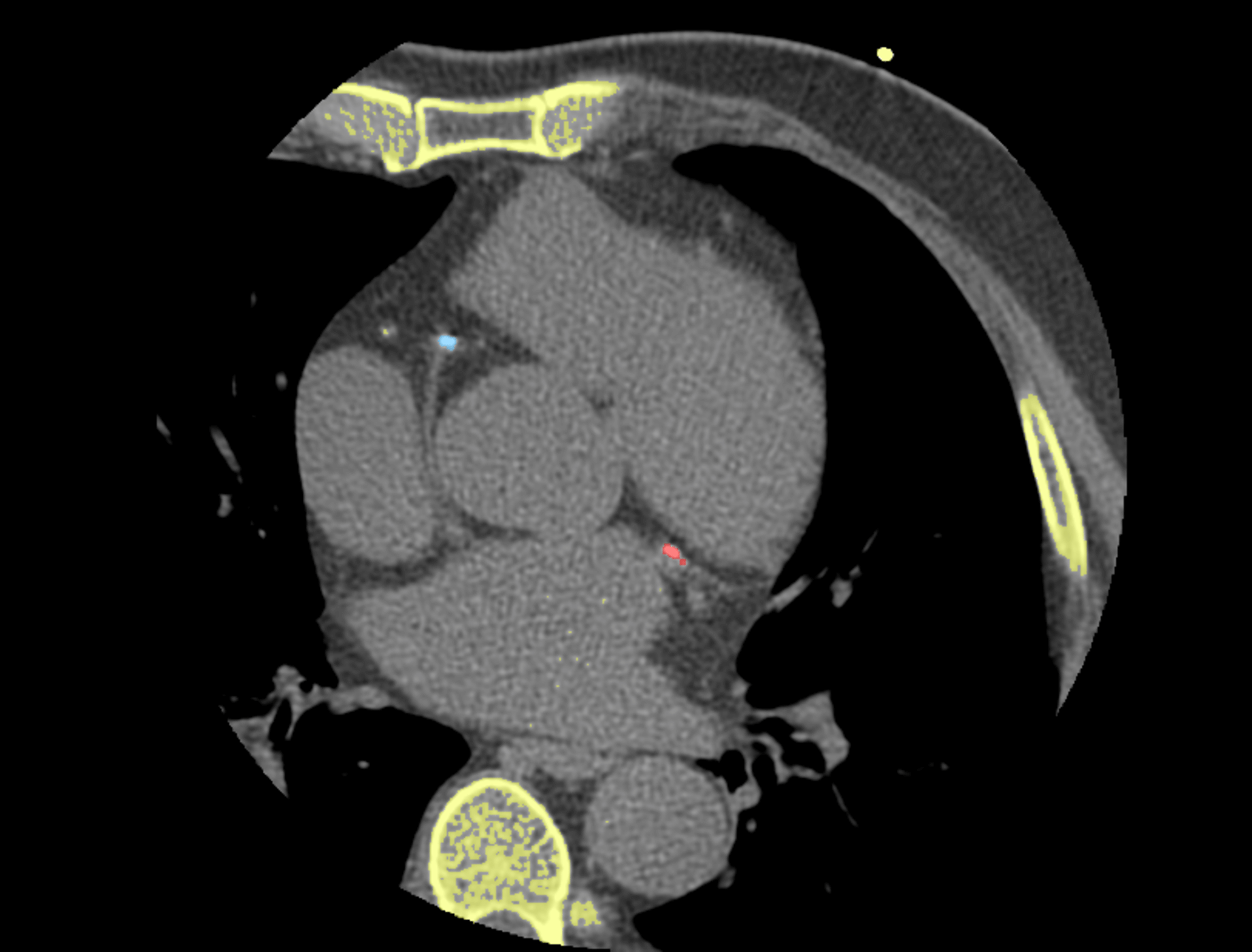
Rapid quantitative assessment of coronary artery calcification, assessment methods includes mass score, agatston score, and volume scores.
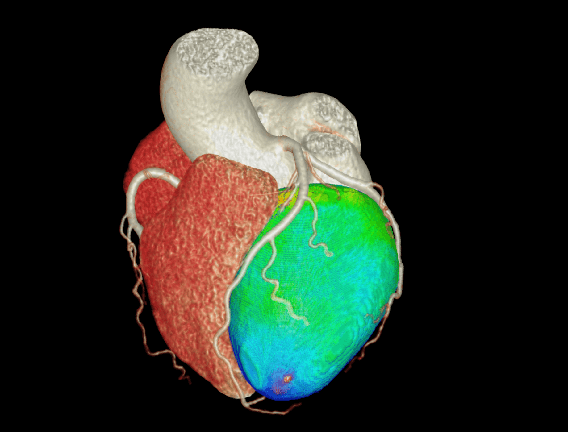
Quickly view the segmented heart and coronary artery, and provide abundant quantitative analysis tools, such as stenosis calculation.
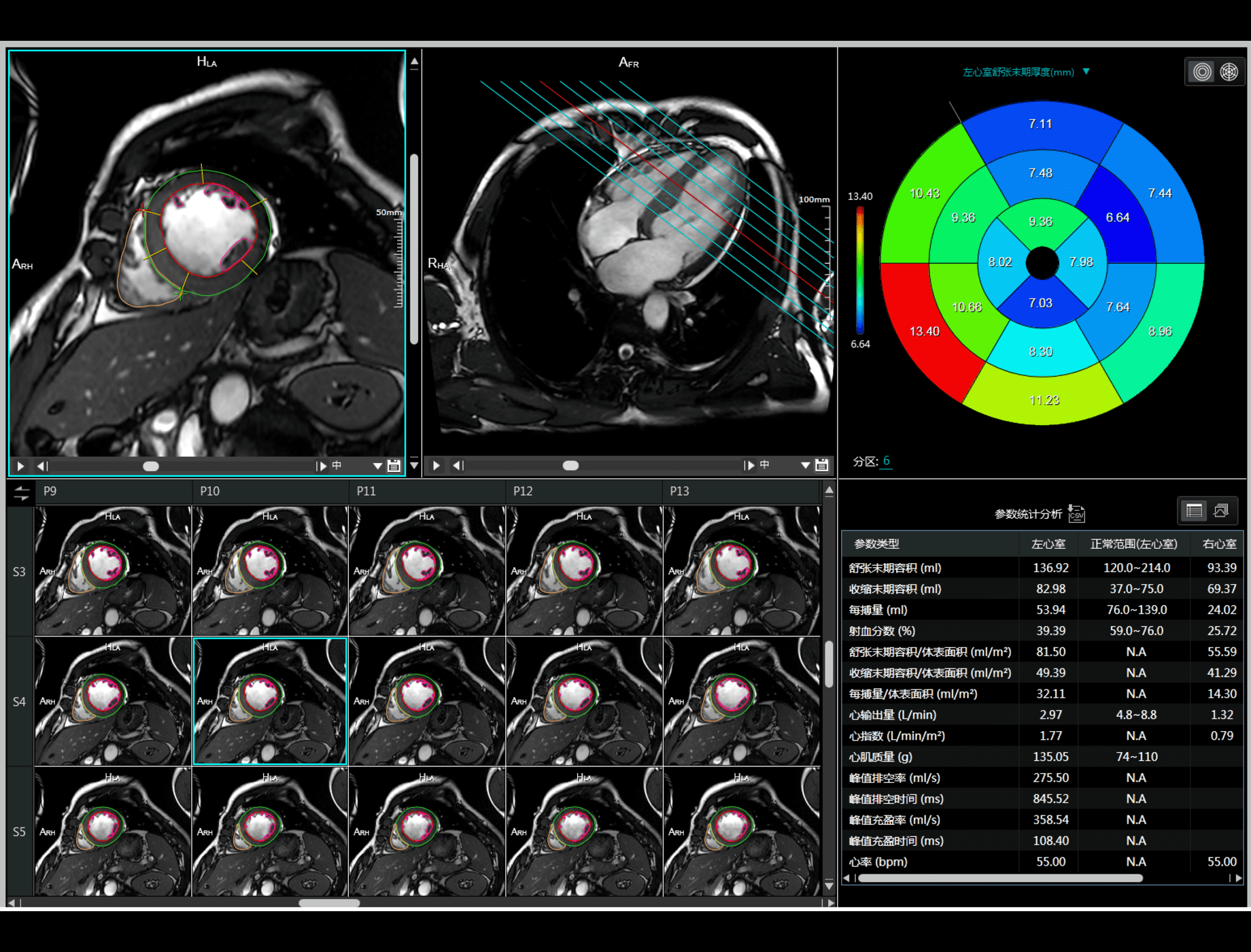
Cardiac Function can complete ventricular function and wall motion analysis automatically. Ejection fraction, cardiac output, wall thickening and other parameters are calculated.
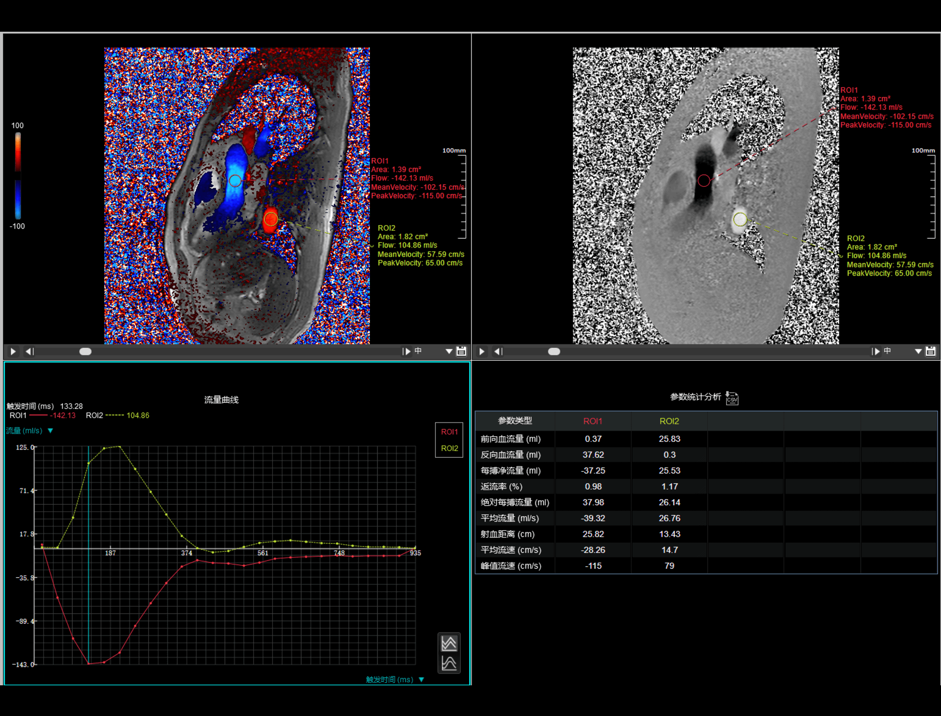
Flow Analysis, based on MR blood flow phase data, calculates velocities, volumes and other information.
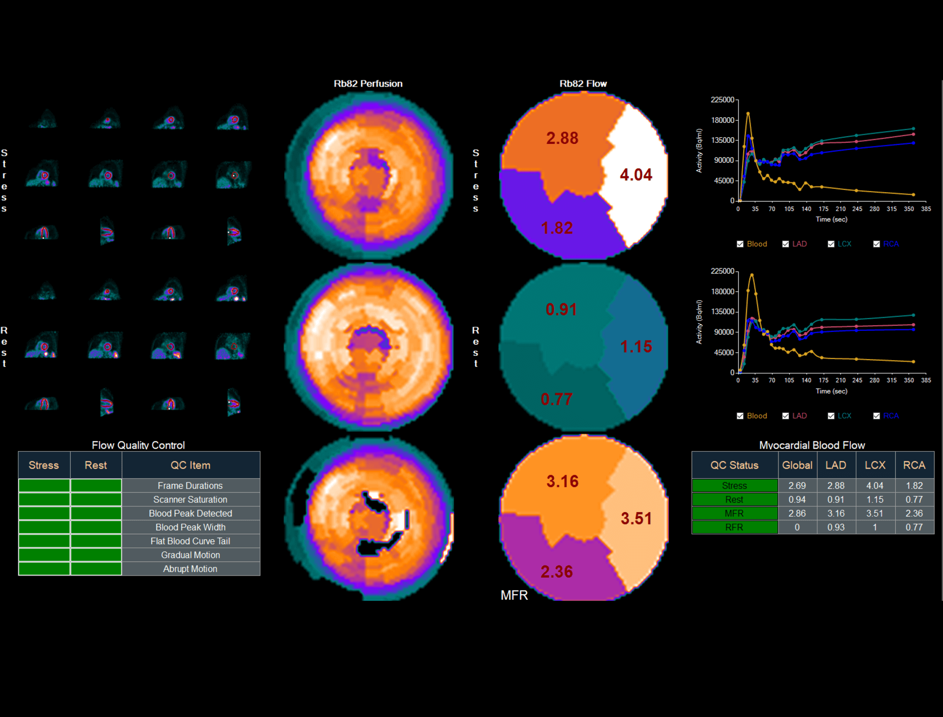
The Cardiac Analysis application provides advanced tools for cardiac SPECT and PET analysis. It supports LV Perfusion Analysis, LV Function parameters calculation, wall thickening evaluation, comparison of perfusion to FDG viability data for mismatch analysis, phase analysis for wall motion, computing myocardial blood flow and myocardial flow reserve from dynamic studies.
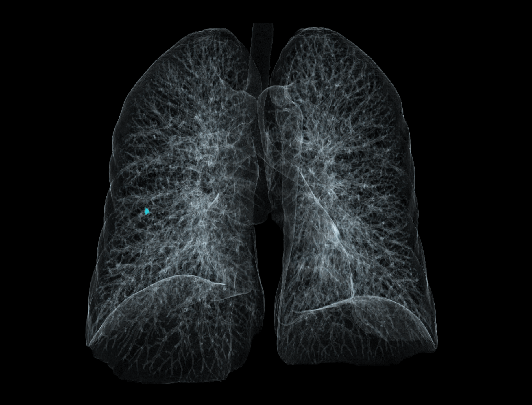
Provide quantitative information about the size and change over time of lung nodules regularly.
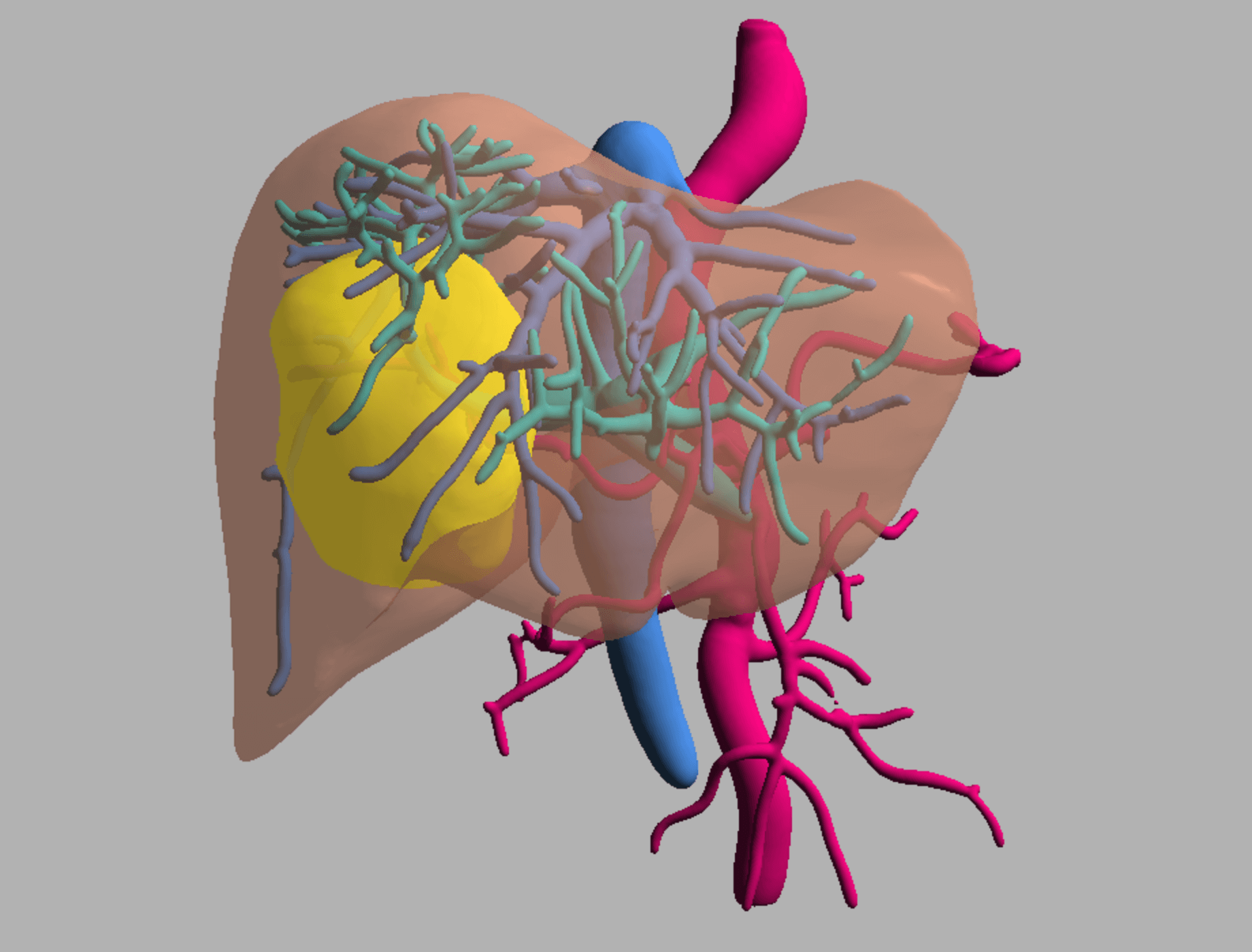
Three-dimensional visualization are offered to analyse relationship between tumor, blood vessel and liver. Support quantitative parameter calculation.
Provide lesion segmentation function, support quantitative statistics of parameters such as volume, change rate, and doubling time of the region of interest in different periods.
Provide perfusion images and ROI tools for rapid assessment of tissue perfusion.
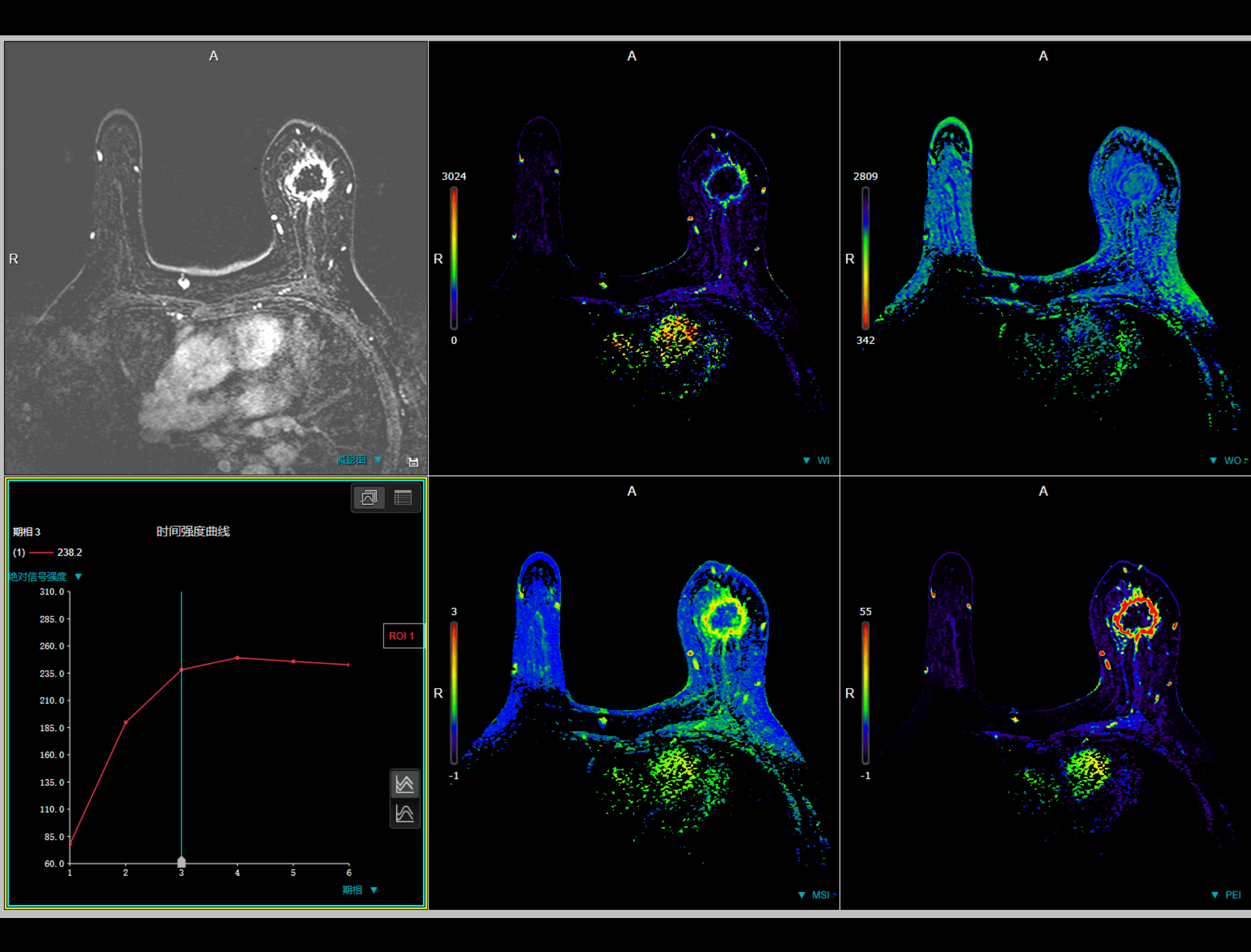
Breast Evaluation is used to analyze enhanced breast MR data and provide parameter maps including Wash-In, Wash-Out, Maximum slope of increasing and Positive enhanced integration ect.
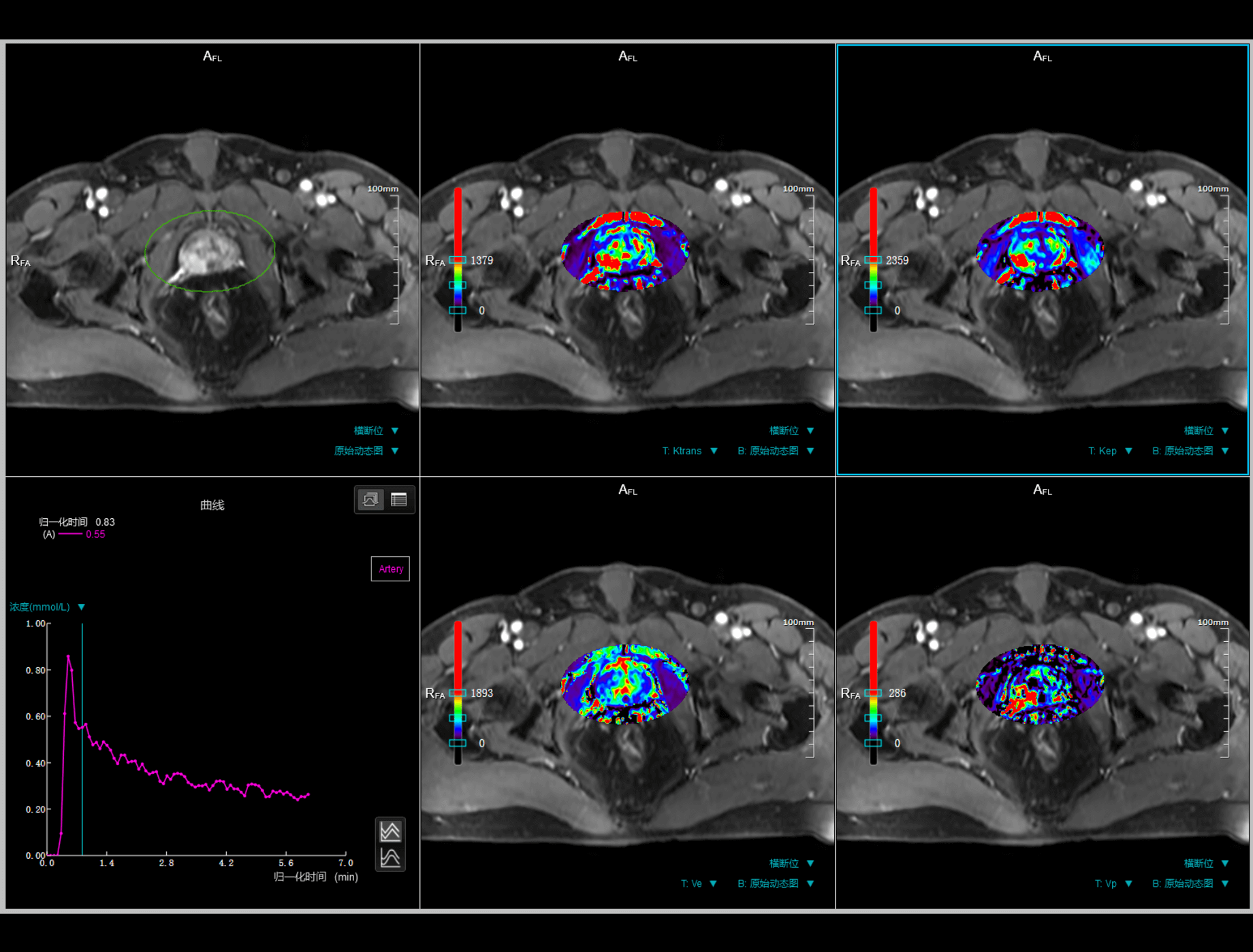
DCE application aims to analyze dynamic contrast enhanacement MR data, can calculate parameters including Ktrans, kep, ve, etc. based on selected pharmacokinetic model.
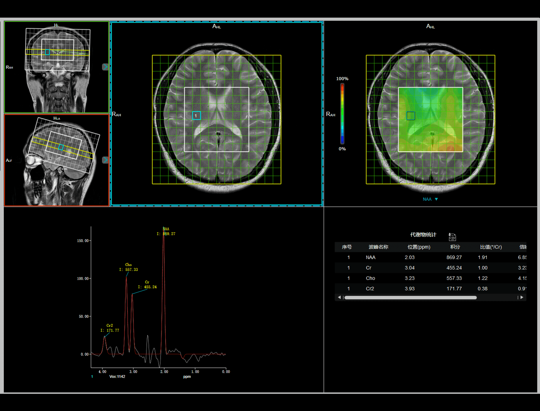
MR Spectroscopy supports the analysis for both SVS(Single Voxel Spectroscopy) and CSI (Chemical Shift Imaging) data for evaluating the molecule constitution and spatial distribution of cell metabolism.

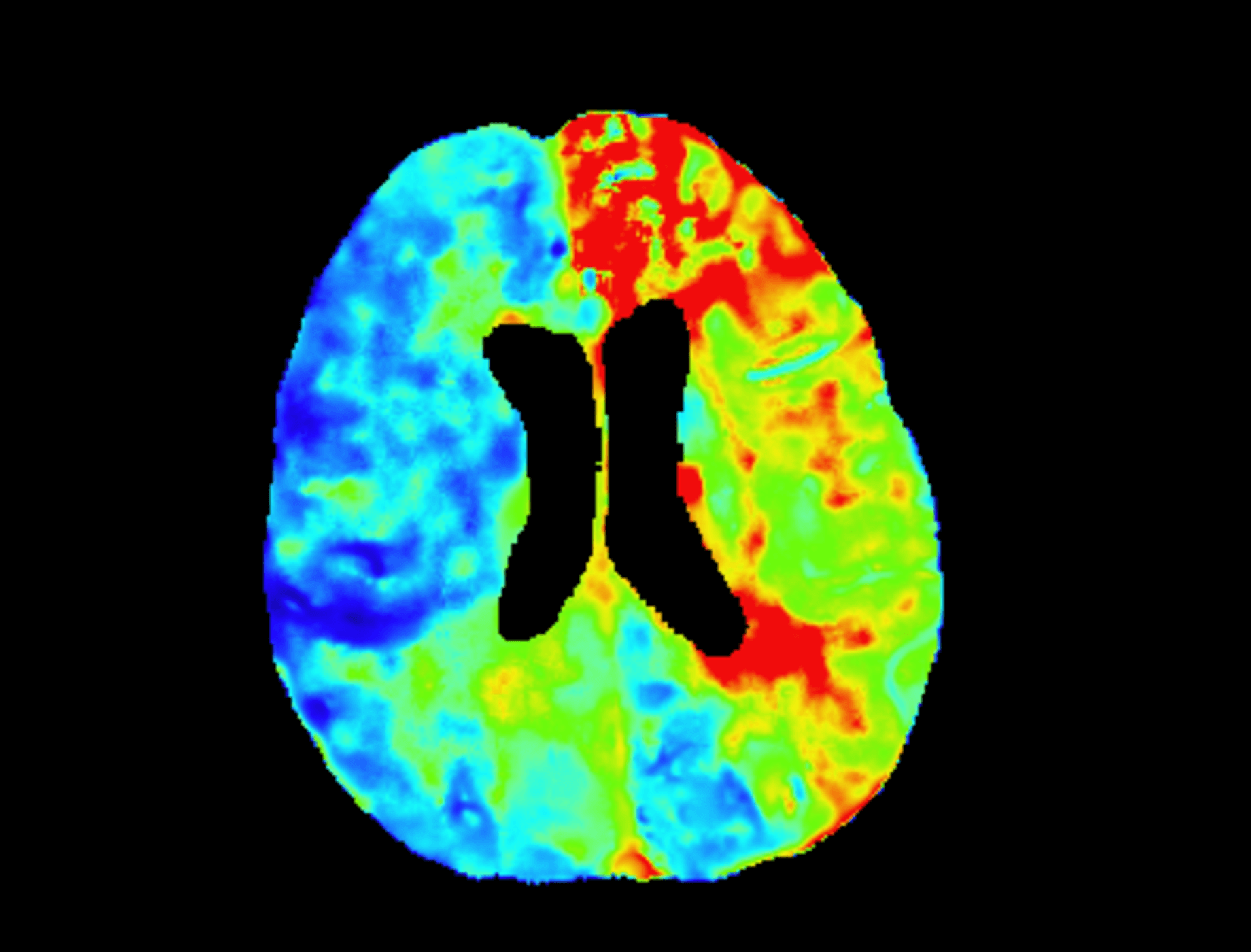
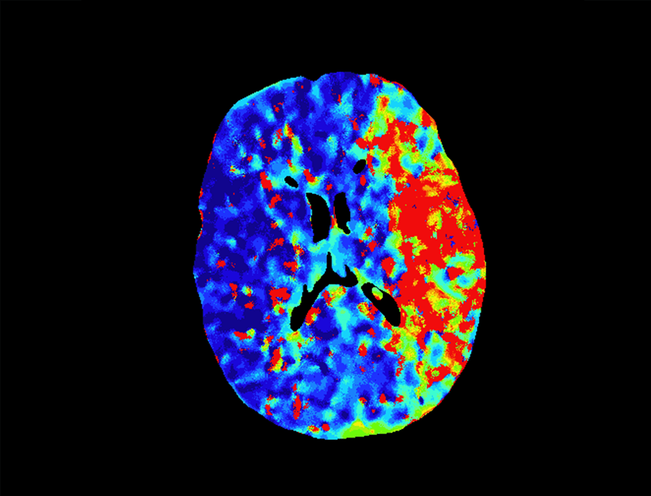
Brain Perfusion able to calculate rCBF (relative Cerebral Blood Flow), rCBV (relative Cerebral Blood Volume), MTT (Mean Transit Time), TTP (time to peak) by different mathematical models, and display corresponding parametric maps.
The application supports compare mode viewing of patient follow-up data, lesion semi-automatic segmentation, and statistical analysis of tumor size and metabolic activity.
Provide perfusion images and ROI tools for rapid assessment of tissue perfusion.
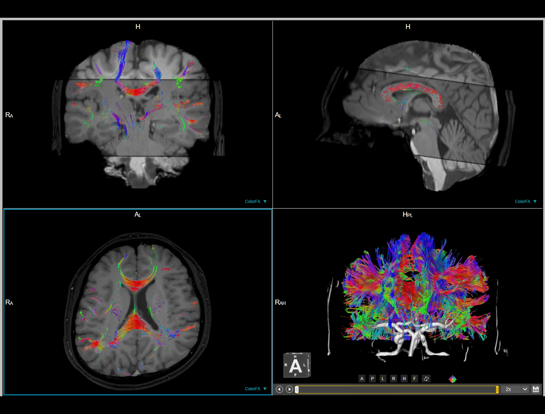
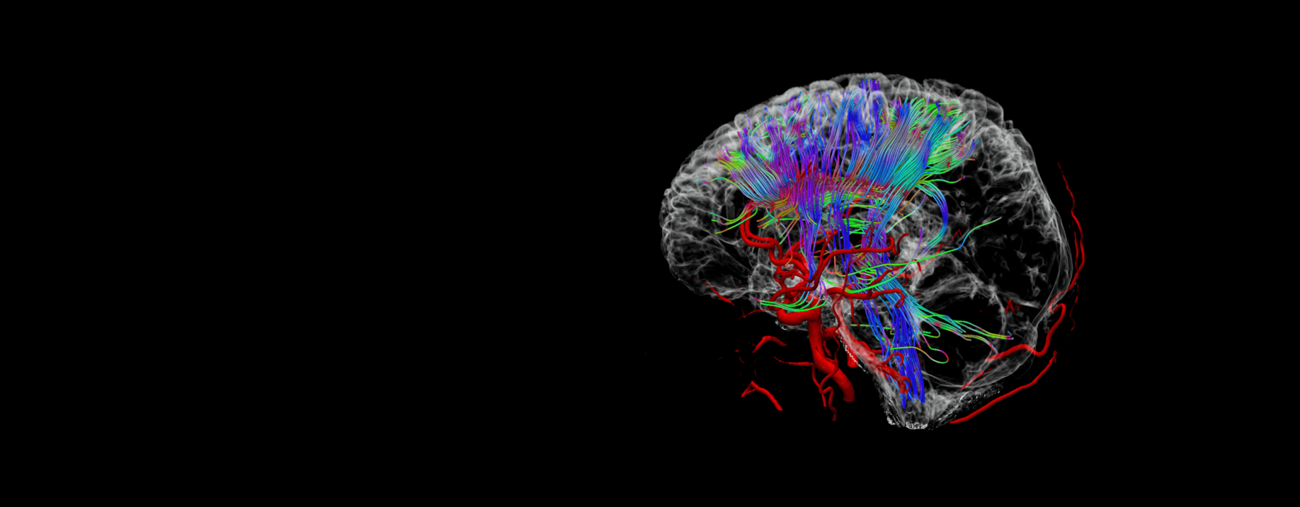
United Neuro is used to analyze functional magnetic resonance imaging (fMRI) and diffusion tensor imaging(DTI) data. According to various fusion and analysis function, users can observe relation among brain function, fiber bundles, lesions and vessels.
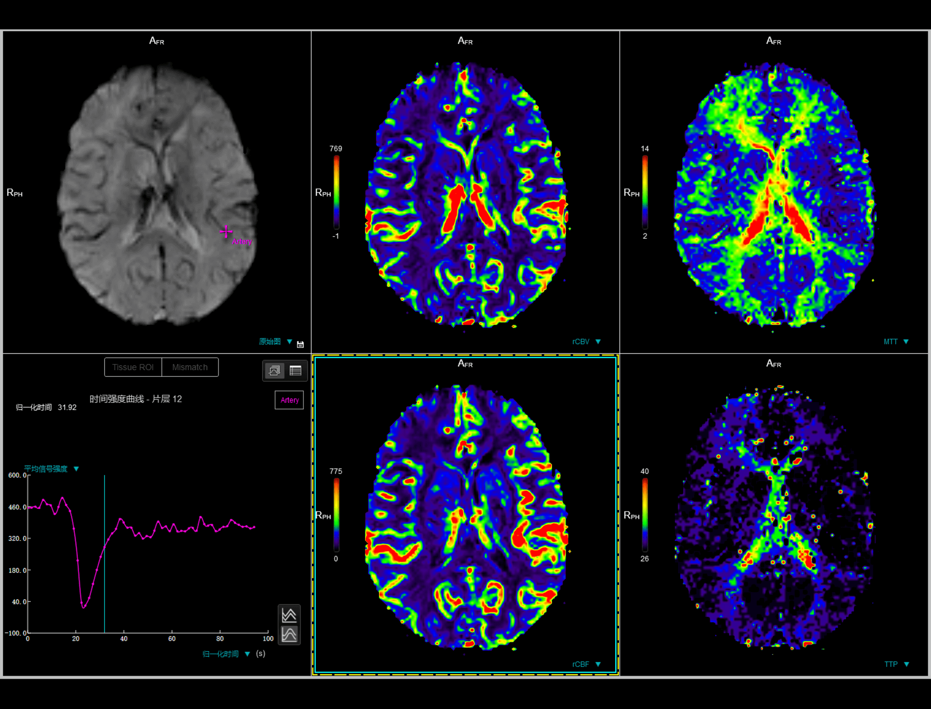
Brain Perfusion able to calculate rCBF (relative Cerebral Blood Flow), rCBV (relative Cerebral Blood Volume), MTT (Mean Transit Time), TTP (time to peak) by different mathematical models, and display corresponding parametric maps.
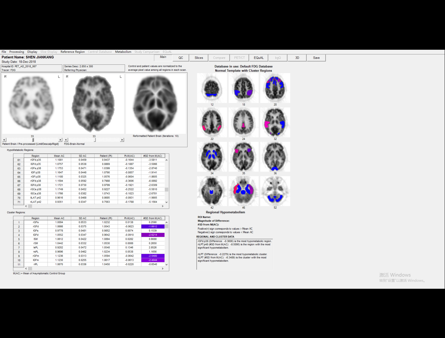
The Brain analysis application is designed to help clinicians perform quantitative analysis by comparing the regional brain activity in an individual scan to that of asymptomatic control subjects for the differential diagnosis of dementia.
Quickly view the vascular structure after bone removal, while providing multi-planar reconstruction and quickly save functions.
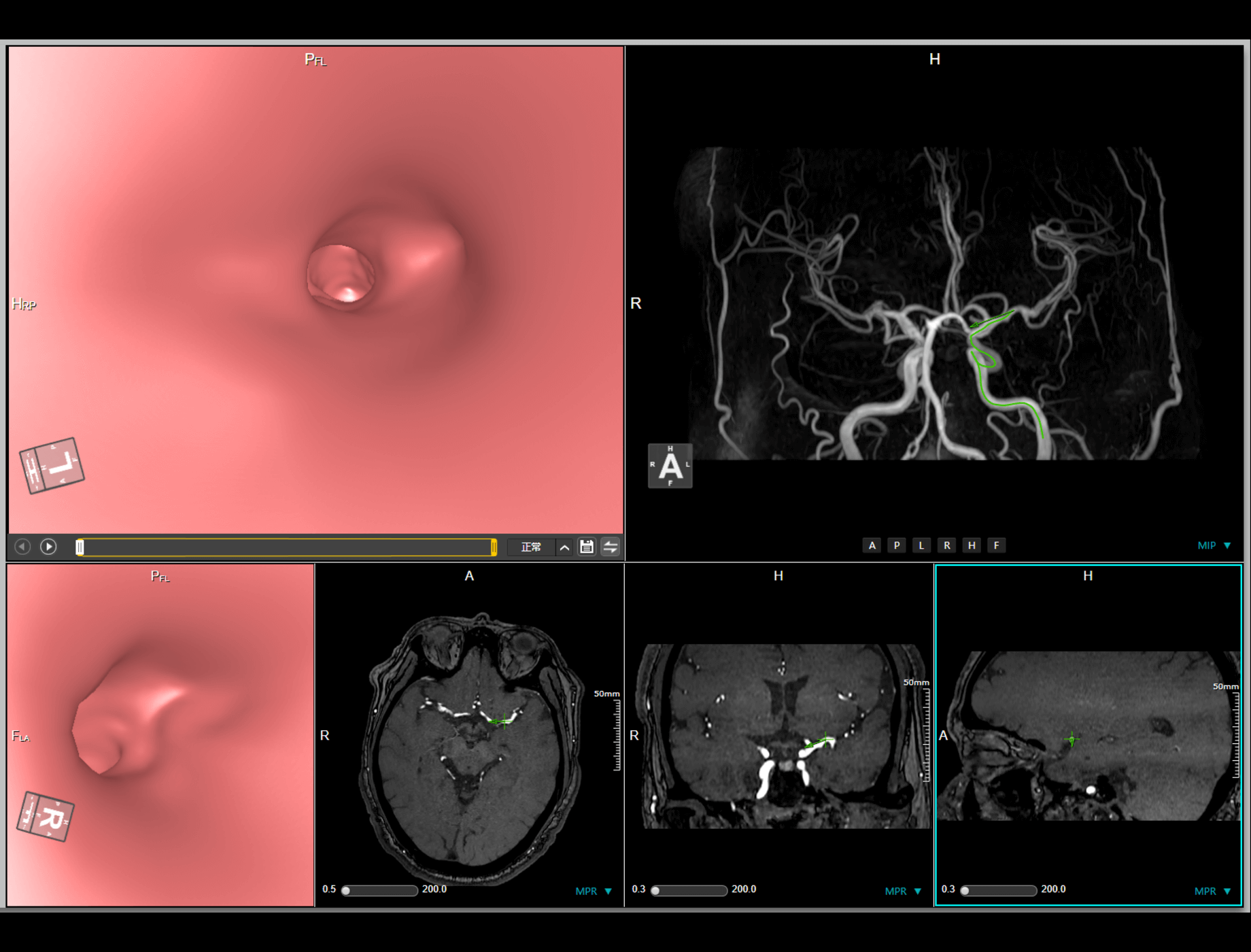
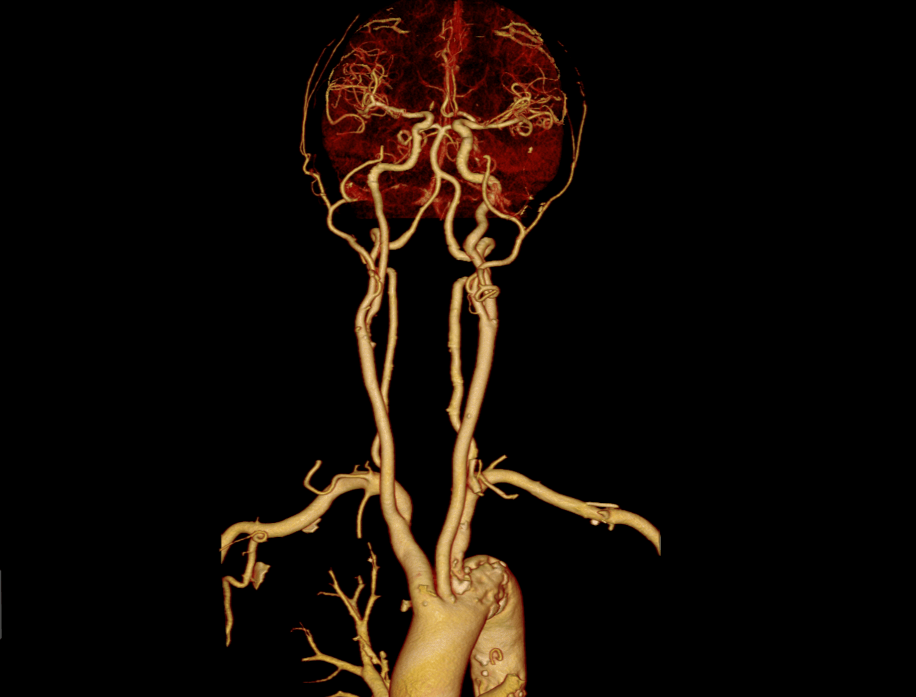
Provide bone removal and blood vessel extraction functions to help observe whether there are occlusions or malformations in intracranial blood vessels.
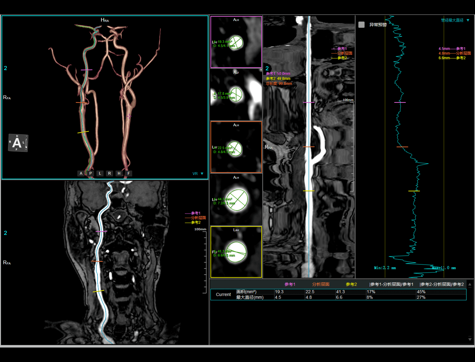
Vessel Analysis provides vessel centerline extraction and contour measurement functions, users can evaluate the stenosis measurements.
Provide virtual endoscopy view to observe the internal structure of the tissue.
Different types of images can be fused and displayed to provide more reference information.
Provide panoramic and cross-sectional views, provide auxiliary reference information for orthodontic treatment plans.
Rapid quantitative assessment of coronary artery calcification, assessment methods includes mass score, agatston score, and volume scores.
Quickly view the segmented heart and coronary artery, and provide abundant quantitative analysis tools, such as stenosis calculation.
Provide quantitative information about the size and change over time of lung nodules regularly.
Quickly view the vascular structure after bone removal, while providing multi-planar reconstruction and quickly save functions.
Provide lesion segmentation function, support quantitative statistics of parameters such as volume, change rate, and doubling time of the region of interest in different periods.
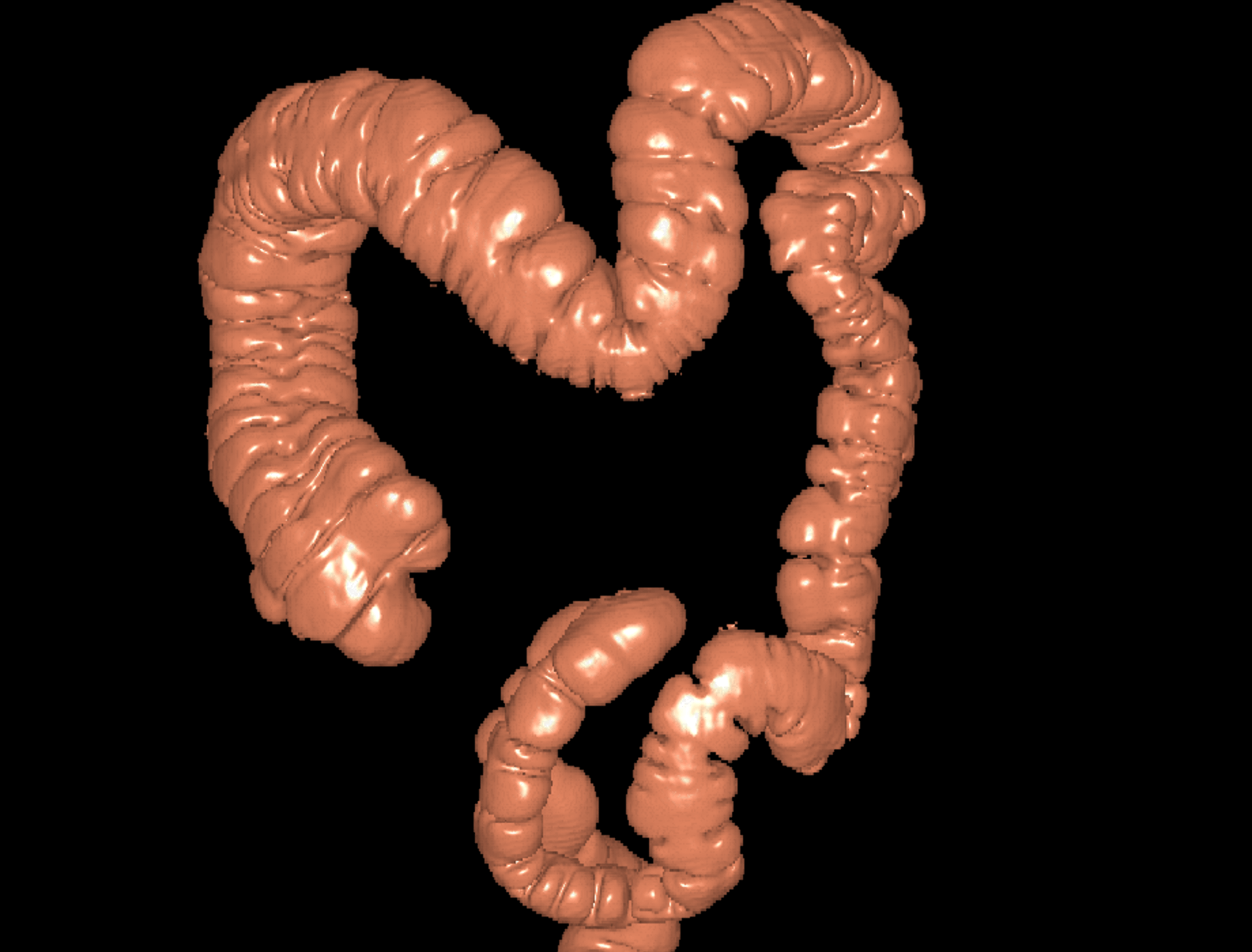
Quickly view the three-dimensional structure of the colon and provide quantitative analysis tools for polyps.
Provide perfusion images and ROI tools for rapid assessment of tissue perfusion.
Three-dimensional visualization are offered to analyse relationship between tumor, blood vessel and liver. Support quantitative parameter calculation.
Provide bone removal and blood vessel extraction functions to help observe whether there are occlusions or malformations in intracranial blood vessels.
Rapid rib segmentation and extraction are offered to assist in rapid fracture positioning.
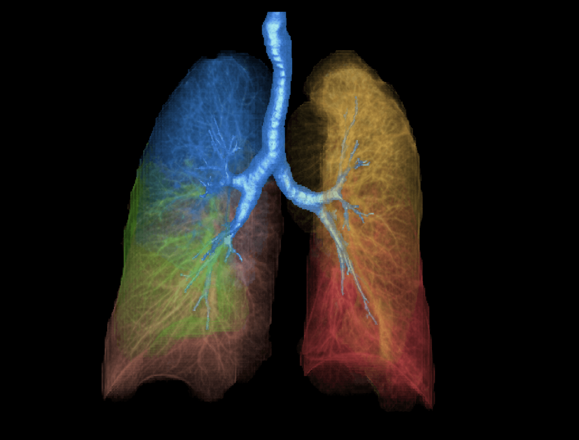
Provide the quantitative parameters calculation and structure information to help evaluate the lung density and airway structure.
Brain Perfusion able to calculate rCBF (relative Cerebral Blood Flow), rCBV (relative Cerebral Blood Volume), MTT (Mean Transit Time), TTP (time to peak) by different mathematical models, and display corresponding parametric maps.
Stitching can stitch several images scanned by segments for panoramic view. Automatic stitching can rapidly gain spine, vessel and whole body stitched images.
MR Spectroscopy supports the analysis for both SVS(Single Voxel Spectroscopy) and CSI (Chemical Shift Imaging) data for evaluating the molecule constitution and spatial distribution of cell metabolism.
Dynamic Evaluation is used to analyze dynamic magnetic resonance images. It provides mean time intensity curve of ROI and parameter maps of positive analysis or negative analysis.
MR image fusion can fuse different images by image registration technology, support to adjust blend ratio,pseudo-color scenarios and reference data.
United Neuro is used to analyze functional magnetic resonance imaging (fMRI) and diffusion tensor imaging(DTI) data. According to various fusion and analysis function, users can observe relation among brain function, fiber bundles, lesions and vessels.
Cardiac Function can complete ventricular function and wall motion analysis automatically. Ejection fraction, cardiac output, wall thickening and other parameters are calculated.
Flow Analysis, based on MR blood flow phase data, calculates velocities, volumes and other information.
Breast Evaluation is used to analyze enhanced breast MR data and provide parameter maps including Wash-In, Wash-Out, Maximum slope of increasing and Positive enhanced integration ect.
Vessel Analysis provides vessel centerline extraction and contour measurement functions, users can evaluate the stenosis measurements.
Inner View offers segmentation algorithms to automatically or manually extract the dividing lines of vessel, and simulates an endoscopy display within the organ by using a 3D display from different angles.
DCE application aims to analyze dynamic contrast enhanacement MR data, can calculate parameters including Ktrans, kep, ve, etc. based on selected pharmacokinetic model.
Image Fusion offers quick and configurable protocols for fused PET/CT and PET/MR image reading and convenient common tools to help you achieve the outcomes you need.
The application provides comprehensive tools for calculating and plotting metabolism activity in a user determined region of interest (ROI) over time and location.
The Brain analysis application is designed to help clinicians perform quantitative analysis by comparing the regional brain activity in an individual scan to that of asymptomatic control subjects for the differential diagnosis of dementia.
The Cardiac Analysis application provides advanced tools for cardiac SPECT and PET analysis. It supports LV Perfusion Analysis, LV Function parameters calculation, wall thickening evaluation, comparison of perfusion to FDG viability data for mismatch analysis, phase analysis for wall motion, computing myocardial blood flow and myocardial flow reserve from dynamic studies.
The application supports compare mode viewing of patient follow-up data, lesion semi-automatic segmentation, and statistical analysis of tumor size and metabolic activity.
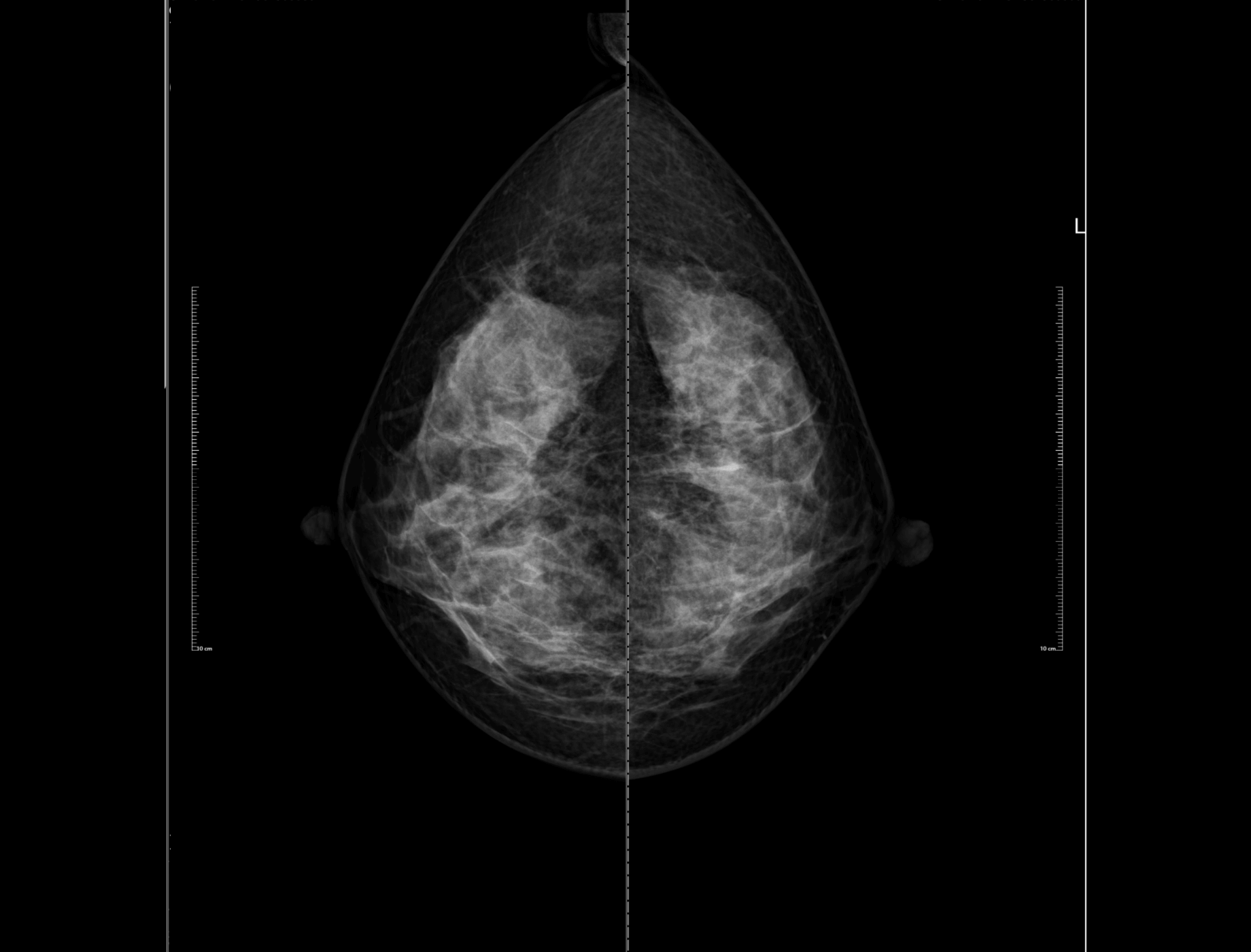
uWS-Mammo supports the display, storage and transmission of both Ultrasound and MR images (DICOM 3.0) on PACS, which are acquired from multiple devices of different vendors.
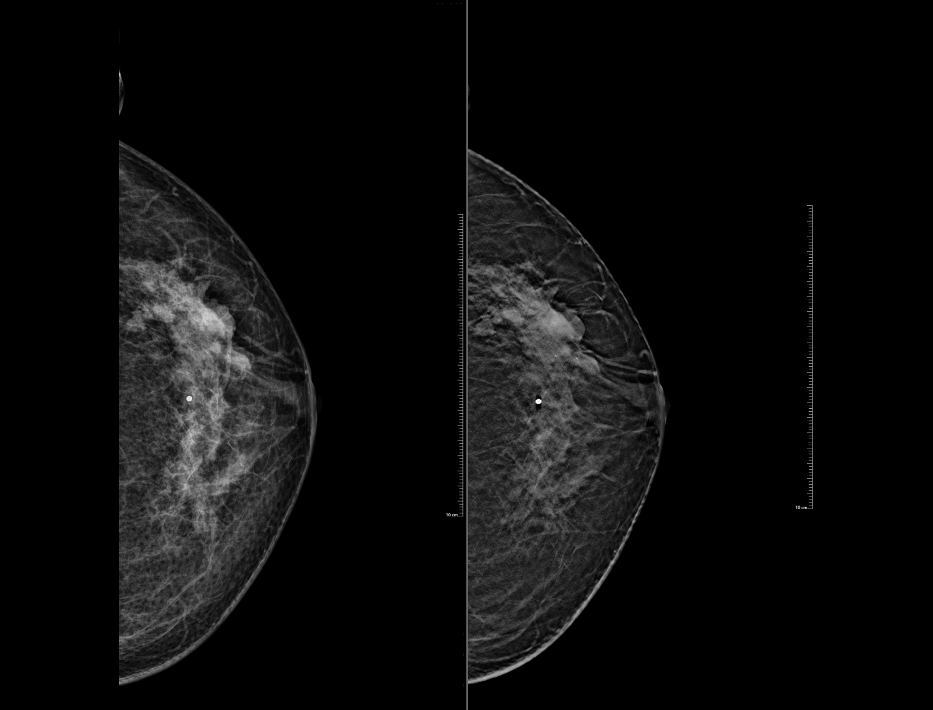
uWS-DBT provides a powerful basis for dedicated mammography screening. It's ideal for viewing standard 2D and tomosynthesis images, film printing, images achiving, report editing, and also supports viewing multi-modality DICOM images.
Industry leading cutting-edge technologies, together with United Imaging's full line of equipment, constitute the underlying core of all software for our products. With good scalability and robustness, that software is one of the core products in United Imaging's mid-end strategy.
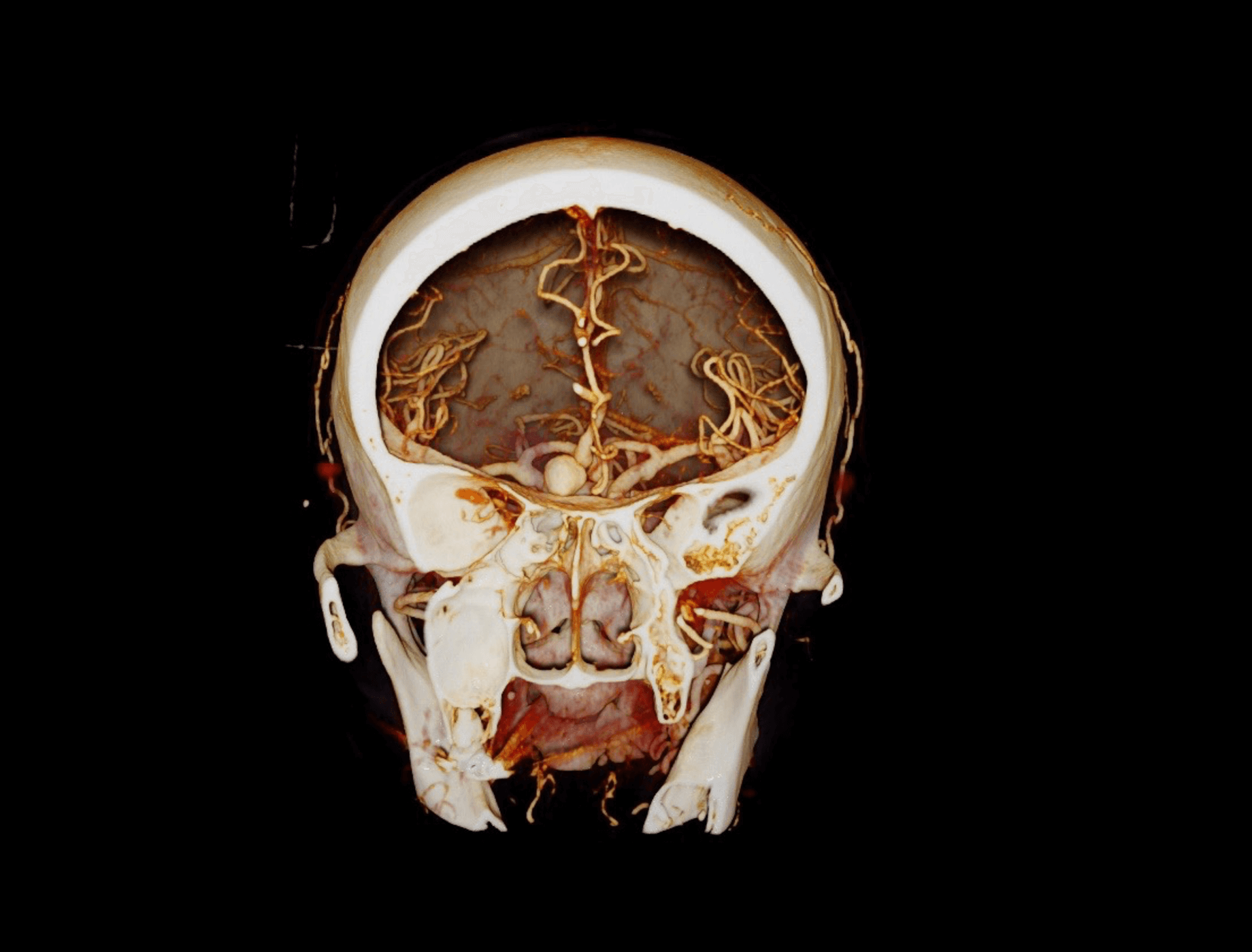
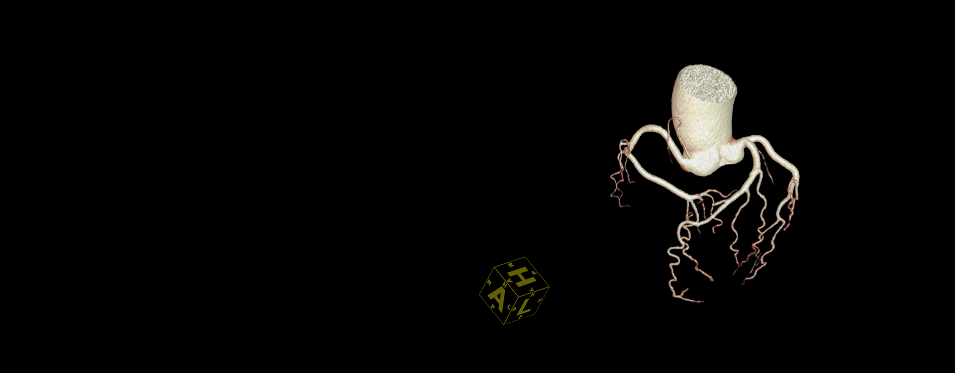
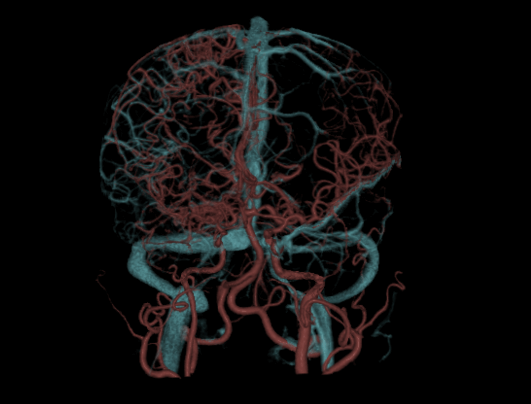
Neuro imaging to accurately display vessel and aneurysm
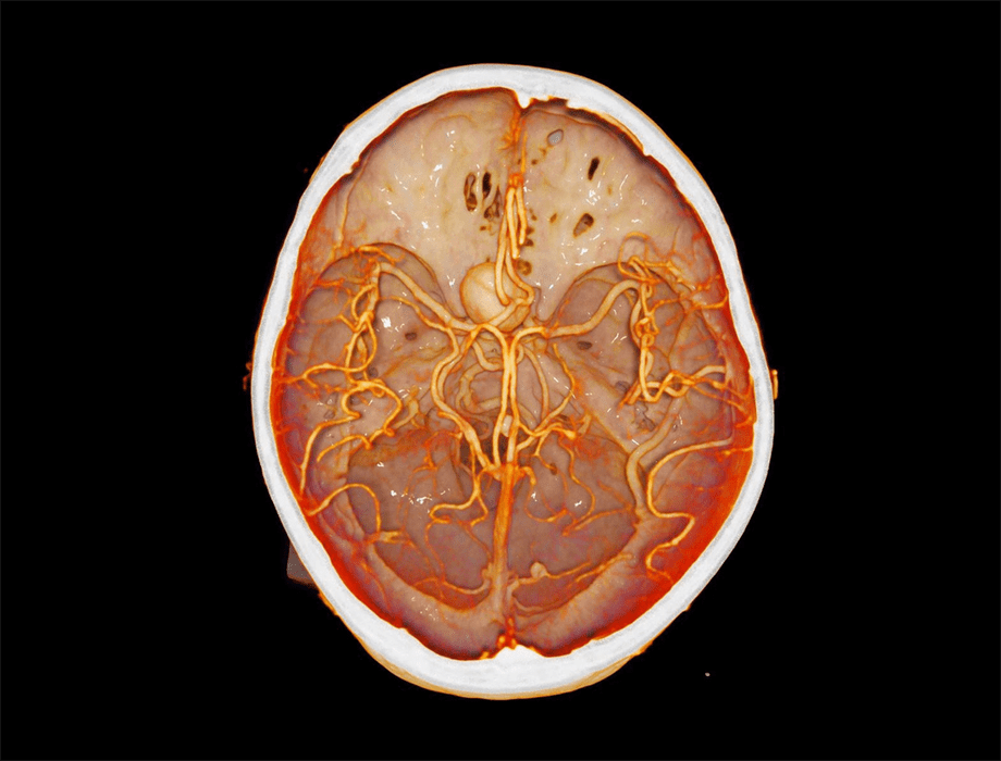
Elegant anatomy HRR volume rendering of intracranial aneurysm and vessel.
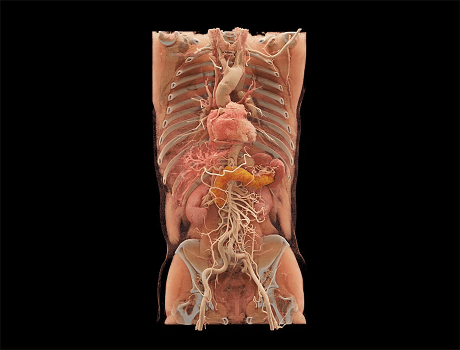
Elegant anatomy HRR volume rendering of whole body organ, bone, vessel and muscle.
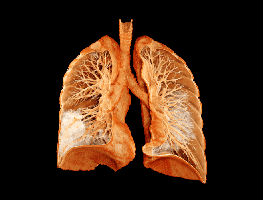
Elegant anatomy HRR volume rendering of pulmonary trachea, vessel and pneumonia.
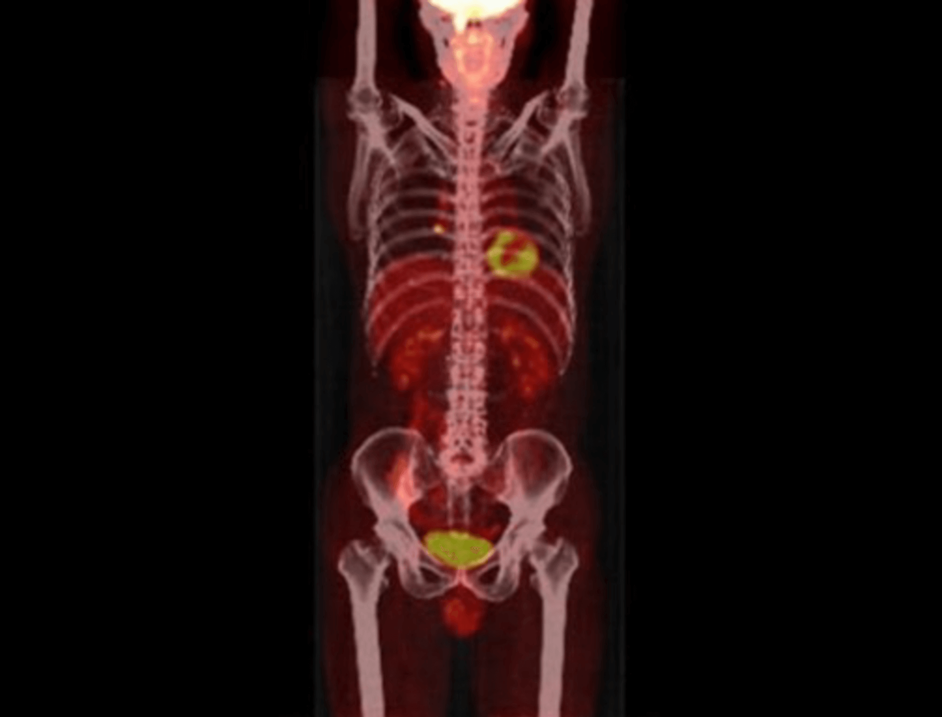
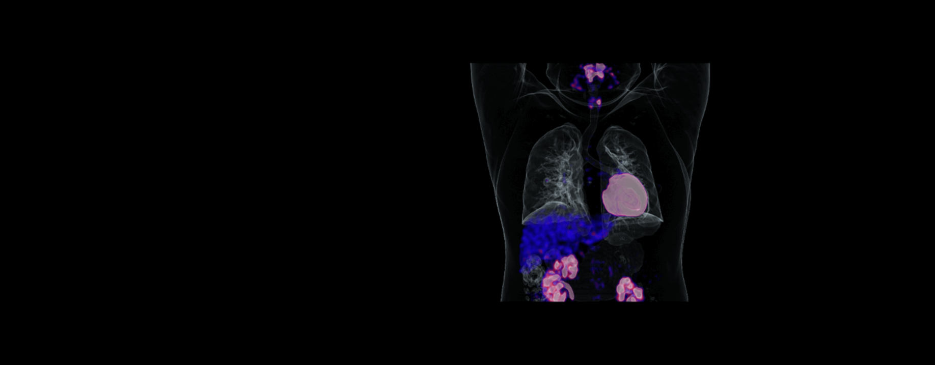
Make auto-registration between whole body CT and PET images.
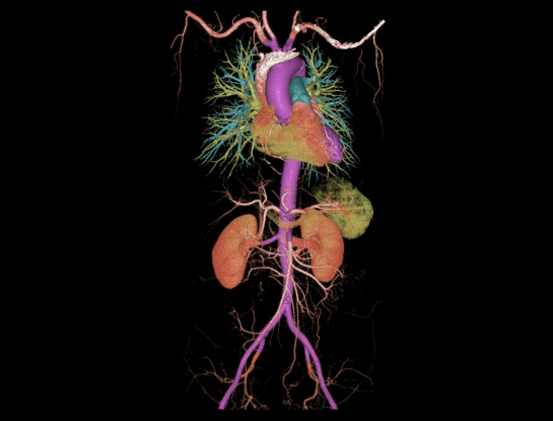
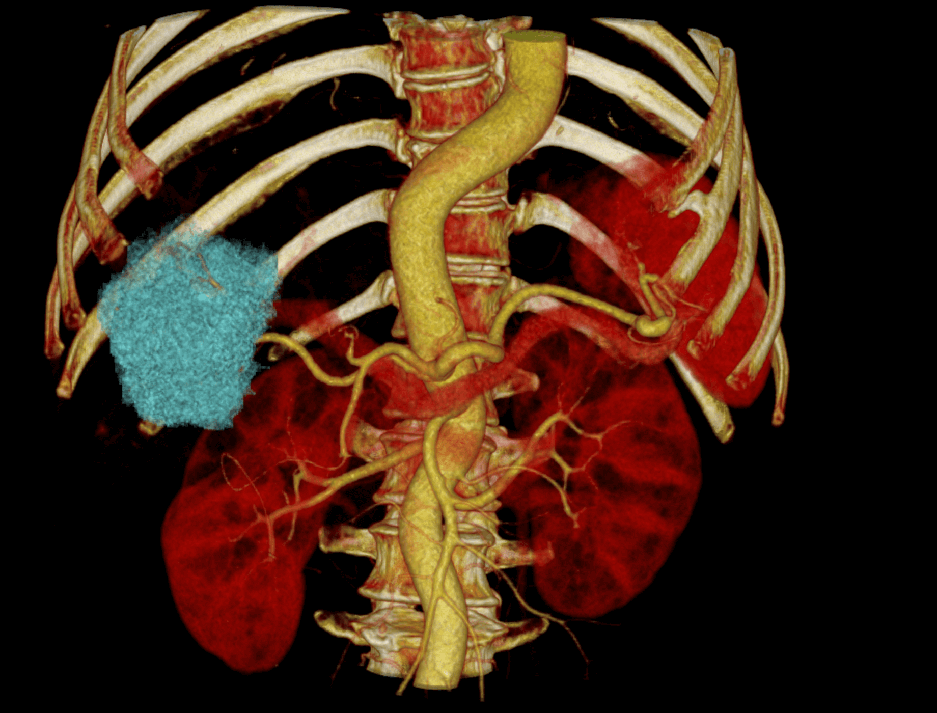
Segment whole body vessel precisely.
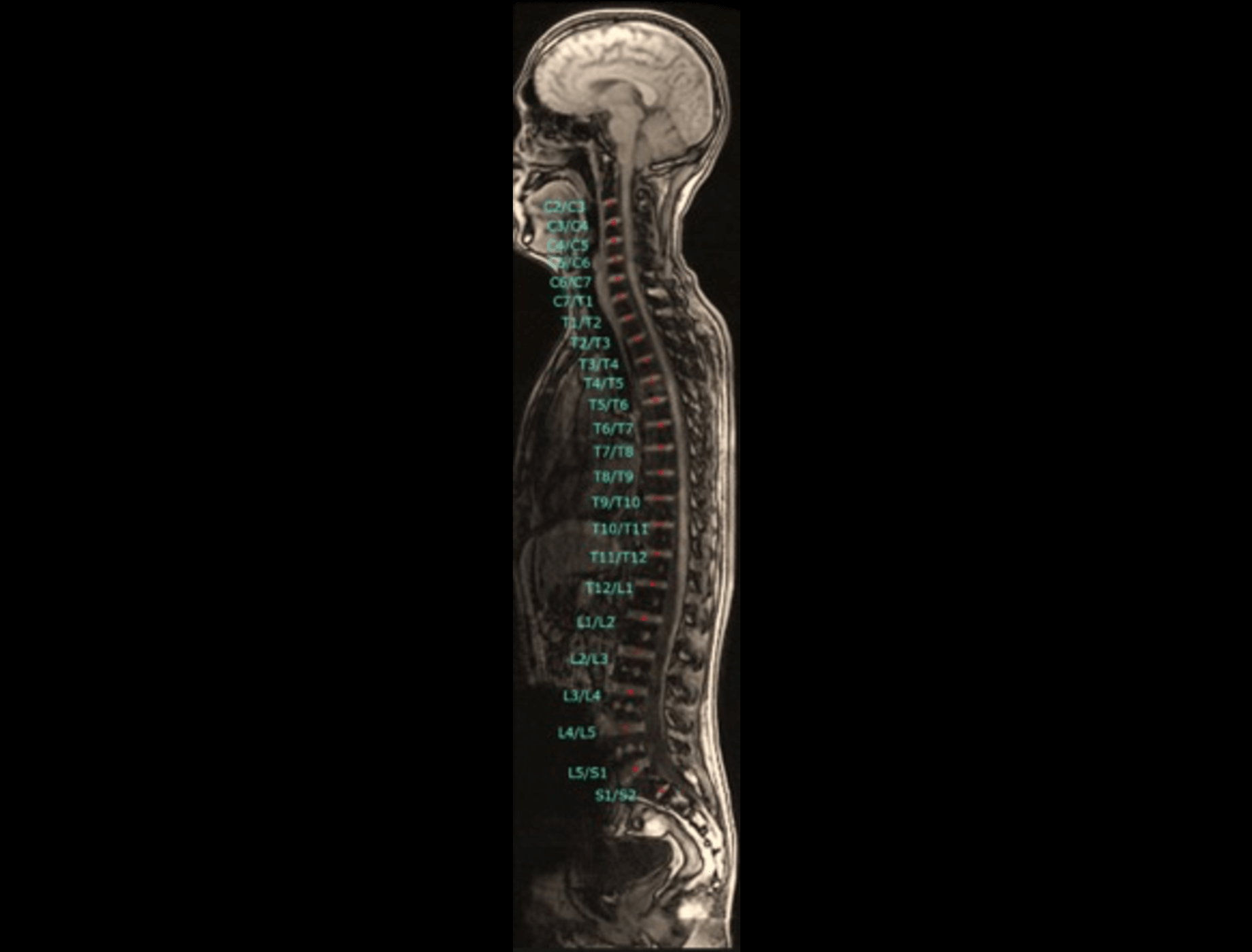
Detect and label vertebral discs efficiently.
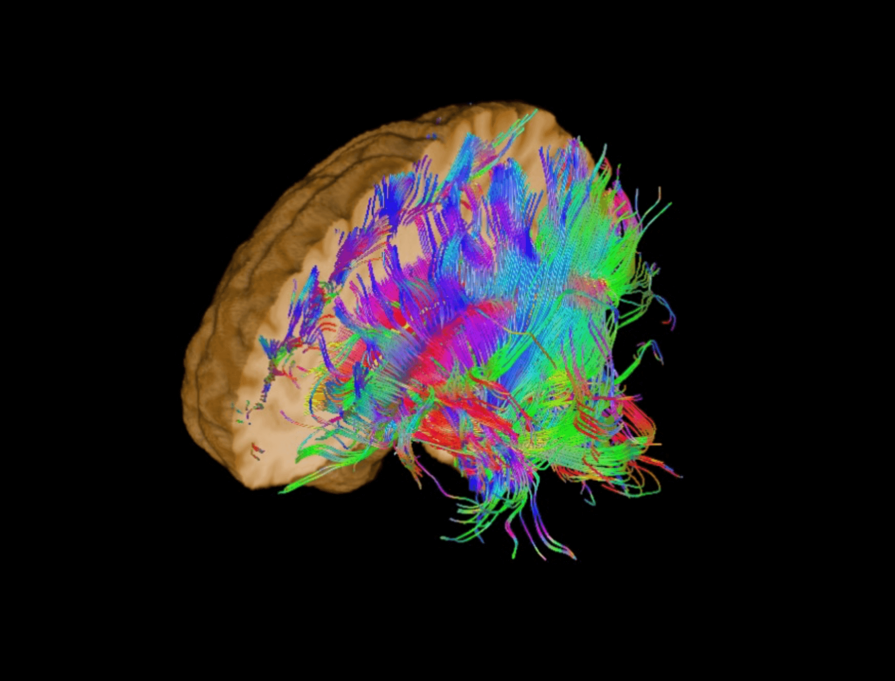
One-stop display of brain parenchyma and fibers tracts.
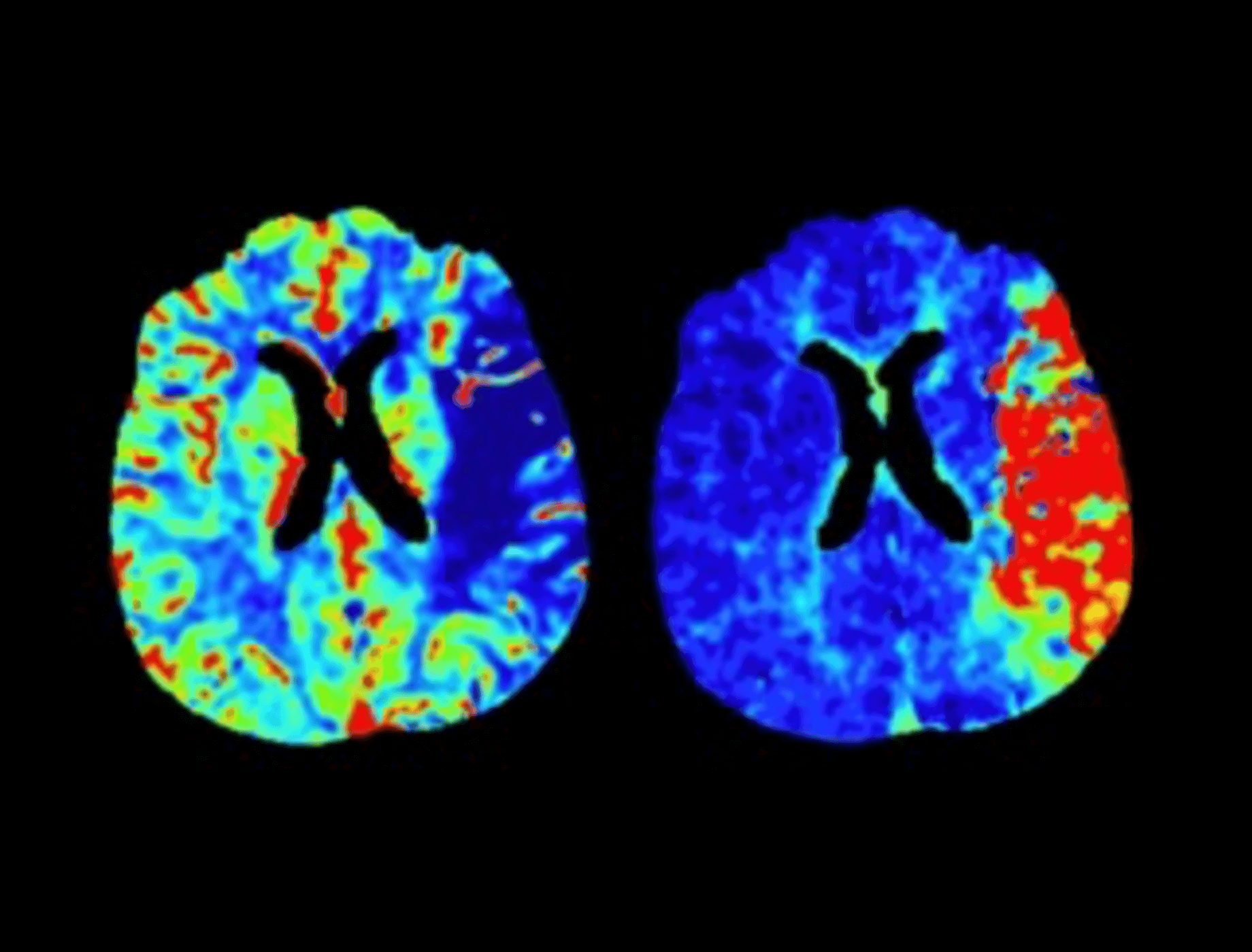
Quantify the degree of cerebral ischemia and infarction of stroke patients.