The shoulder and shoulder girdle are complex structures that may sustain many different types of injuries. A shoulder CT scan makes it possible to diagnose all kinds of abnormalities occurring in the shoulder joint, involving both bones and soft tissues, muscles, joints, ligaments or veins. Importantly, it is a quick, painless and non-invasive examination, which allows precise diagnosis and monitoring of therapeutic effects.
The shoulder girdle is a complicated anatomical structure consisting of two bones – the clavicle (collar bone) and the scapula (shoulder blade). This structure connects the upper limb to the torso. The clavicle is connected with the scapula via the acromioclavicular joint (AC joint); it is also connected with the sternum via the sternoclavicular joint (SC joint). The scapula, on the other hand, is connected to the arm bone via the glenohumeral joint (shoulder joint). Therefore, the shoulder girdle plays a key role in arm mobility and stability. Because of its complex structure and wide range of motion, the shoulder is susceptible to a variety of injuries. A shoulder scan performed using modern CT scanners is an examination that allows precise 3D imaging of this structure.
Indications for a shoulder CT scan include:
Computed tomography of the shoulder is a radiological imaging examination that provides extremely detailed results, which are very helpful in diagnosing all kinds of injuries and conditions. A CT scan of the shoulder and shoulder girdle allows accurate evaluation of the bones, joints, muscles, tendons and other soft structures within the shoulder joint. It is particularly useful in detecting injuries such as ruptures, fractures and rotator cuff tendon damage as well as in evaluating inflammation and degenerative conditions. CT scans of the shoulder can also be used to assess treatment progress after injuries or surgeries performed in this area.
Therefore, a shoulder CT scan is applicable in:
- injury diagnosis – detecting fractures, ruptures as well as ligament, tendon and muscle injuries
- assessment of degenerative conditions – analysis of conditions such as arthrosis
- analysis of lesions – detection of tumours or other abnormalities within the shoulder area
- postoperative diagnosis – evaluation of surgical treatment effects
- detection of inflammatory lesions and abscesses – analysis of inflamed areas within the shoulder joint
- evaluation of congenital anomalies – examination of congenital defects of the shoulder
- shoulder prosthesis (arthroplasty) assessment – aseptic or septic glenoid loosening
Shoulder CT scan – is a doctor’s referral required?
A CT scan of the shoulder requires a referral from a doctor. Because this examination involves X-rays, it cannot be performed without medical supervision. For this reason, even if a patient intends to undergo a shoulder CT scan at a private medical facility, he or she must have a doctor’s referral.
While United Imaging Healthcare’s state-of-the-art CT scanners only emit minimal radiation doses as they are able to achieve extremely accurate imaging at low doses thanks to their innovative detection systems and software that utilises artificial intelligence algorithms, and thus make the examination safer, there is still some radiation exposure.
What does computed tomography of the shoulder look like?
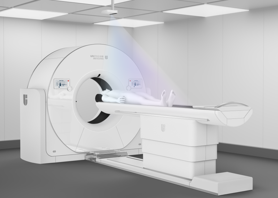
For CT scans of the shoulder, there are several possible patient positions during the examination. The first is the traditional supine position. Another is abduction with external rotation – the arm is bent at the elbow with the hand resting behind the head. Yet another position is adduction with internal rotation when the patient touches the other shoulder with his or her hand. The prone position is possible as well. Depending on the type of diagnostics, the examination may involve several positions. In this case, the patient is asked to reposition his or her arm in a specific manner during the shoulder CT scan.
Computed tomography of the shoulder with contrast
A shoulder CT scan with contrast allows even more accurate diagnosis of the shoulder, especially in terms of assessing the condition of vascular structures. This examination is referred to as CT angiography of the shoulder. This type of imaging test is usually recommended when the following conditions are suspected:
- vascular anomalies, such as changes in the structure of blood vessels or aneurysms
- congestion and blood clots that can lead to tissue ischaemia
- vascular tumours
- internal bleeding
At most seven days before a CT scan of the shoulder with contrast is conducted, creatinine levels must be tested to check the condition of the kidneys. The administration of contrast also involves additional preparations before the examination. First of all, the patient should be fasting. Moreover, the administration of contrast is associated with the risk of allergic reaction. If this has happened before, the patient should inform the doctor and medical staff. Hyperthyroidism and obvious allergy to the contrast agent are also contraindications to contrast administration.
However, in the case of previous allergic reactions, special medications are administered to minimise the possibility of recurrence. Nevertheless, performing a CT scan of the shoulder with contrast must be carefully considered by the doctor, and the patient must be prepared for this examination in all respects.
*ATTENTION! The information contained in this article is for informational purposes and is not a substitute for professional medical advice. Each case should be evaluated individually by a doctor. Consult with him or her before making any health decisions.