Digital imaging, like the invention of X-rays earlier, was a revolution in medical diagnostics, enabling increasingly precise and rapid analysis. This technology goes significantly beyond the capabilities of traditional radiological methods, offering incomparably better image quality and greater diagnosis efficiency.
Digital imaging in medicine
Digital radiological examinations have become a mainstay of modern medical diagnostics, since owing to its advanced capabilities, digital radiography makes it possible to obtain much more accurate images of the human anatomy, finding application in different fields of medicine – primarily dentistry, orthodontics, orthopedics, oncology, but also many others.
Types of digital X-ray examinations:
- Digital radiography (DR) – a direct form of digital radiography, in which images are captured using special digital detectors. This allows images to be obtained immediately, which significantly reduces examination time and increases patient comfort.
- Intraoral digital radiography (radiovisiography) – an imaging method used in dentistry, which allows X-ray photographs to be taken directly in the patient’s mouth, providing detailed and clear images of dental structures and surrounding soft tissues.
- Dental panoramic radiograph (orthopantomogram) – a panoramic digital X-ray showing the jaws, teeth and mandible together with the temporomandibular joints and maxillary sinuses, mainly used in orthodontics and oral surgery and also to detect pathologies in the oral cavity.
- Cephalogram – a digital X-ray of the skull in the lateral, posterior-anterior or axial position, used primarily in orthodontics, for the diagnosis of cranial malformations or to assess skeletal age.
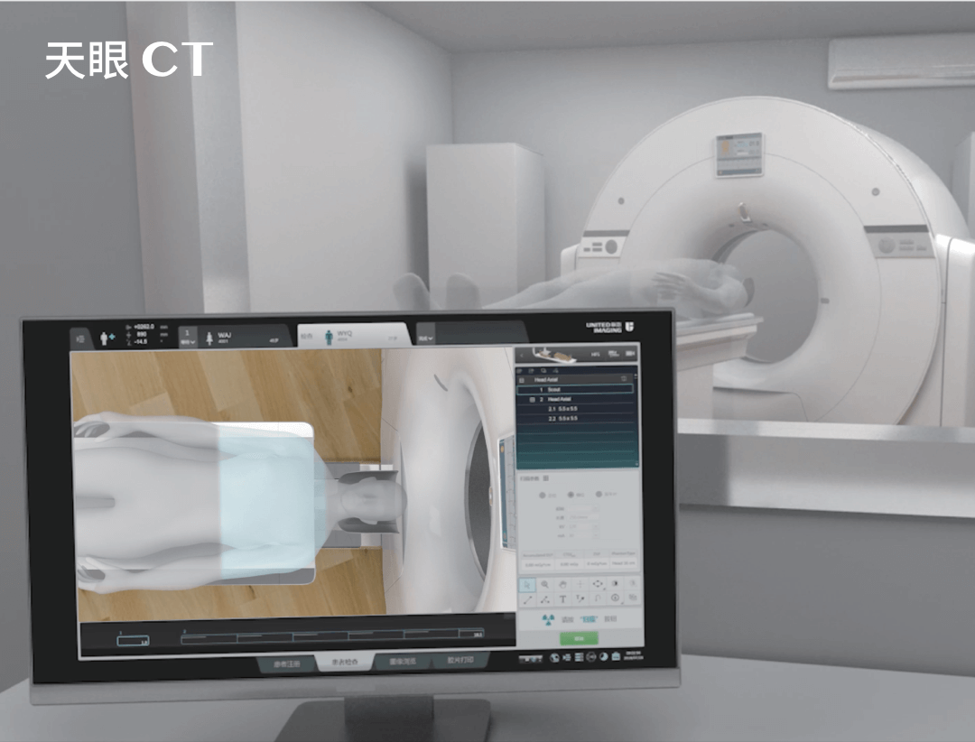
- Computed tomography (CT) – CT scanners use digital radiography to produce detailed cross-sectional images of the human body.
- Digital mammography – a specialised form of digital radiography used to diagnose and monitor breast diseases. It offers high quality images, which is particularly important for detecting early stages of breast cancer.
- Digital fluoroscopy – provides moving images of the inside of the body, which is useful in diagnosing problems with the digestive, vascular or musculoskeletal systems. Digital fluoroscopy enables body functions to be observed dynamically in real time, and is often used in interventional procedures.
Digital radiological diagnostics – history
The field of digital radiology was pioneered in the 1980s by Francis Mouyen, a French dentist from Toulouse who introduced and developed the concept of digital radiography, especially in dentistry. Mouyen developed RadioVisioGraphy (RVG) – the first commercially available digital dental radiography system, which revolutionised the way in which dental X-ray examinations are performed and analysed, paving the way for the development of modern digital imaging techniques in various fields of medicine.
Digital vs. analogue X-ray diagnostics
Digital radiography works on a similar principle to traditional radiography, but instead of on special film, images are created electronically. During the examination, X-rays pass through the patient’s body and are recorded by digital image detectors. These detectors convert the X-rays into a digital signal, which is subsequently processed and displayed on a computer screen.
Digital X-ray diagnostics differs from its analogue version in image quality, efficiency and speed of data processing. It allows doctors to obtain much more detailed and clearer images of internal body structures, which significantly improves diagnosis accuracy and treatment effectiveness.
Another significant advantage is that digital images can be easily transmitted, stored and processed, which is impossible with traditional techniques. In addition, the use of digital technologies makes it possible to significantly reduce the radiation dose used during the examination.
Digital imaging applications – digital radiography in practice
Digital radiography is very widely used in many areas of medicine, including bone and joint, lung and heart imaging as well as dental diagnostics and mammography.
Digital X-rays open a wide variety of analytical possibilities, enabling:
- contrast and color saturation to be modified
- precise measurements to be taken
- spatial images to be generated
- cross-sectional images of the density of specific structures to be generated
The ability to obtain fast and precise results makes digital imaging particularly useful in situations that require immediate diagnosis, such as traumatic injuries or acute conditions. Thus, it is difficult to imagine modern medicine without digital radiography.
At the same time, the field develops all the time, and an excellent example of this are the modern DR systems offered by UIH, with innovative software and digital solutions that make it possible to obtain precise images with extremely low radiation doses.
*ATTENTION! The information contained in this article is for informational purposes and is not a substitute for professional medical advice. Each case should be evaluated individually by a doctor. Consult with him or her before making any health decisions.