Computed tomography – indications, what does it look like, how to prepare for the scan and how long does a CT scan take?
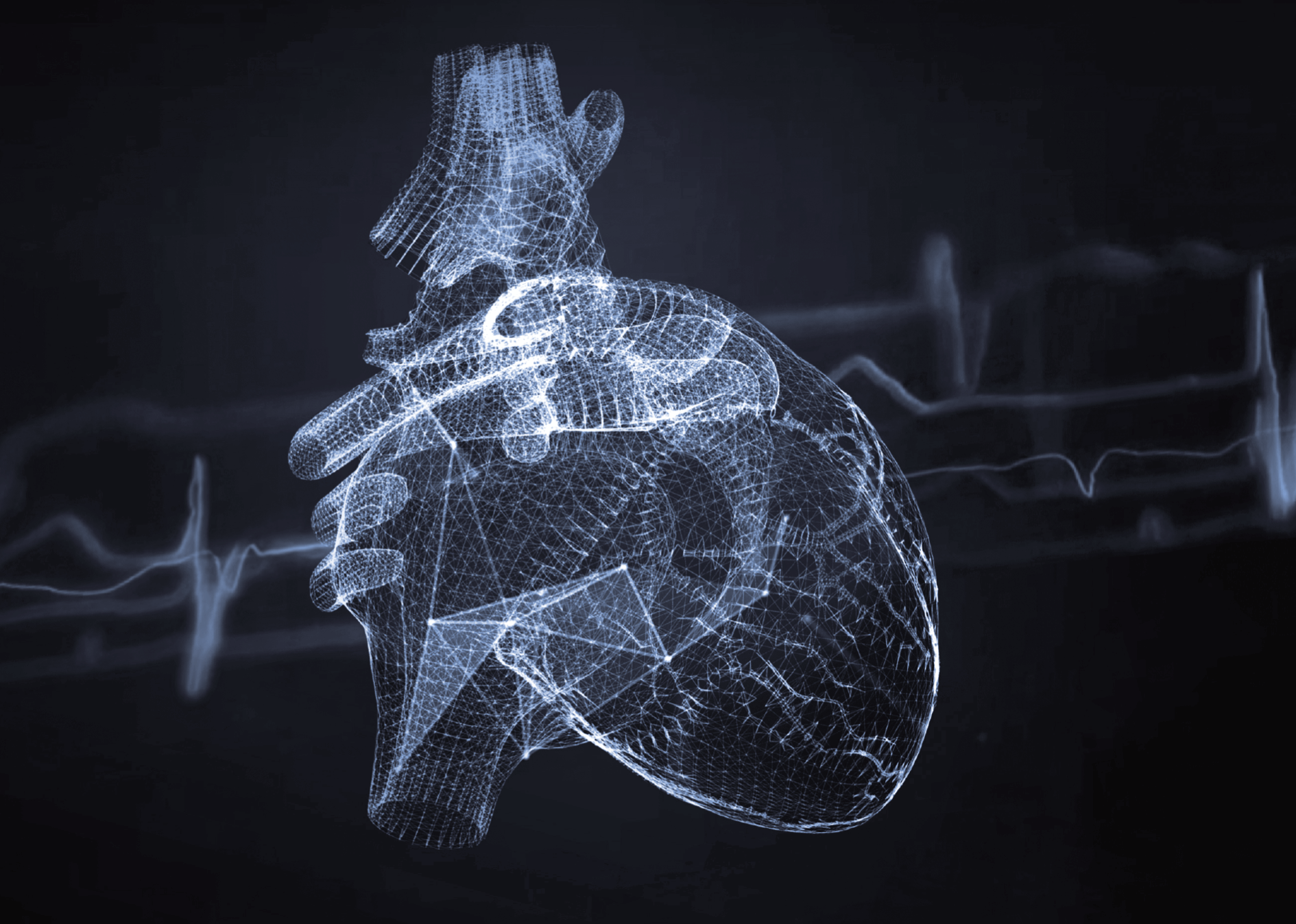
Computed tomography, or CT for short, is one of the greatest advances in radiological diagnostics. By combining X-rays with state-of-the-art equipment and information technology, it produces precise images of internal organs and bones, providing the detailed and accurate data needed to make a proper diagnosis.
What is a CT scan?
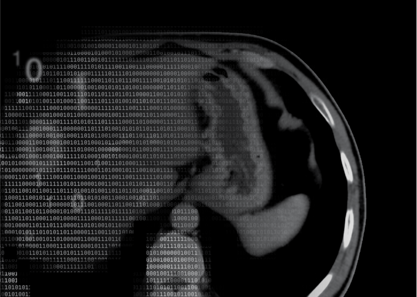
A CT scan is non-invasive, which is one of the major advantages of this imaging modality. During a CT scan, the patient lies on a moving table that slides into the CT scanner. The machine emits X-rays that pass through the body and are picked up by detectors. Because different tissues absorb radiation to different degrees, it is possible to obtain detailed images of the body’s internal structures.
An important aspect of CT is the ability to reconstruct images in different planes, allowing a more accurate assessment of the condition of the structures being scanned. This enables doctors to better understand complex conditions and plan treatment accordingly. As a result, CT scans have become an invaluable tool in the diagnosis of a wide range of conditions, from trauma to cancer.
Origins and development of computed tomography
The history of computed tomography (CT) dates back to the 1960s when Godfrey Hounsfield, an engineer at Electric and Musical Industries Ltd. in the UK, developed a prototype of the first CT scanner. Hounsfield’s revolutionary research contributed to a major breakthrough in medical imaging. The first CT scanner was developed in 1968 and was mainly used for brain diagnostics. The imaging technique was developed in response to the limitations of traditional X-rays, which could not provide sufficiently detailed images of the body’s internal structures.
In 1971, Hounsfield unveiled his invention to the medical world, ushering in a new era of diagnostic imaging. In 1979, Hounsfield was awarded for his achievements in the field of computed tomography the Nobel Prize in Physiology or Medicine, which he shared with Allan Cormack. In the years that followed, CT developed as a tool for examining the brain, as well as other parts of the body, including the chest, abdomen and extremities. The use of multi-row detectors in modern CT scanners has reduced examination time and improved image quality.
The development of CT technology has been driven by the need to produce more accurate images of the body’s internal structures. Today’s CT scanners use advanced data analysis software to produce 3D image reconstructions, allowing doctors to better understand complex anatomical structures, which is essential for effective diagnosis and treatment planning.
Indications for CT scans
Computed tomography is one of the most commonly performed imaging tests. It is the first choice for diagnosing head and spinal injuries, as well as suspected intracranial haemorrhage or brain abscess. Head CT is also used to diagnose brain tumours, cerebrovascular disease and skull bone disease.
How do I prepare for a CT scan?
Preparation for a CT scan varies depending on the type of scan, but the most common guidelines are as follows:
- Fast for at least 6 hours before the scan
- Limit fluid intake 3 hours before the scan
- Remove metal objects such as jewellery or watches
- Inform the medical staff of any allergies, pregnancy, claustrophobia, and medication you are taking
- If you are diabetic, it is recommended that you take insulin and eat a meal before the scan
In the case of an abdominal scan, you may be asked to drink an aqueous contrast agent solution to help visualise internal structures. It is also important to empty your bladder before the scan.
CT scans with and without contrast – differences
There are two types of CT scan - with and without contrast. Contrast-enhanced scans involve the administration of a contrast agent to improve the visibility of internal organs. Contrast is given intravenously or orally and is particularly helpful in diagnosing cancer, inflammation and vascular disease. Non-contrast scans are less invasive and do not require the administration of additional substances, but images may be less detailed in some cases.
CT scan – step by step
A CT scan involves the following steps:
- The patient lies down on a moving table
- The table slides inside the gantry of the machine where the X-rays generated by the tube pass through the patient’s body
- Images are recorded in the transverse plane and then reconstructed in three dimensions
- The patient is released from the gantry
How long does a CT scan take?
The duration of a CT scan depends on the type of scan being performed and can range from a few to several minutes. More complex examinations may take longer. It should be noted that the patient should cooperate with the medical staff in order to make the examination as effective as possible.
Contraindications to CT scans
The main contraindications to CT scans are pregnancy and allergy to the contrast medium. CT scans should also not be repeated frequently because they involve exposure to X-rays.
The characteristics of CT scans make them an invaluable diagnostic tool. Most importantly, CT allows for very rapid and accurate diagnosis, which is essential for proper treatment planning, especially in emergencies.
*ATTENTION! The information contained in this article is for informational purposes and is not a substitute for professional medical advice. Each case should be evaluated individually by a doctor. Consult with him or her before making any health decisions.