What does contrast enhancement in CT mean?
Contrast enhancement in computed tomography (CT) scans refers to a technique used during a CT scan whereby a contrast agent is administered to the patient, allowing certain structures in the patient’s body to become more clearly visible on CT images. How does this work in practice? What are the types of contrast agents and when are they used? Read on to find out more!
Purpose of contrast agent administration
Modern diagnostic medicine makes very frequent use of various kinds of imaging, which primarily involve computed tomography (CT) and magnetic resonance imaging (MRI), since these types of examinations allow very detailed imaging of the body’s internal structures. In some cases, contrast enhancement is used whereby special contrast agents are administered to the patient in order to obtain even more precise images.
Thus, the primary purpose of contrast agent administration is to improve the quality of images obtained during a CT scan. These agents are specifically designed to ‘illuminate’ specific structures or areas in the human body, allowing for more accurate imaging and identification of potential health conditions.
In practice, contrast enhancement involves administering a special contrast agent to the patient, usually containing iodine in the case of CT scans, or gadolinium (so-called paramagnetic agents) in the case of MRI scans. This contrast agent is then distributed throughout the patient’s body. Due to their properties, these substances are clearly visible in CT images, creating contrast between different tissues and structures.
Diagnostic application of contrast enhancement
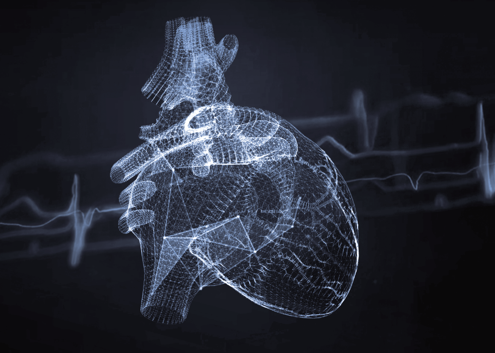
Contrast agents are extremely important in medical diagnostics, since they allow doctors to image structures and tissues that may be difficult to identify using standard CT scans. For instance, if cancer is suspected, a contrast agent may enable the tumour to be more clearly visualised, allowing the doctor to assess its size, location or possible metastases with greater accuracy. In the case of heart disease, contrast agents can help visualise blood vessels and evaluate blood flow, which is of crucial importance to identifying problems such as arterial stenosis, occlusions or myocardial damage.
Examinations in which contrast agents are used
Contrast agents are used in many different CT scans. They can help detect, diagnose and evaluate a variety of conditions and diseases, including cancer, stroke, heart disease, hemorrhage, kidney disease, liver disease, inflammation and infection, nervous system disease and many other pathological changes. Contrast agents can be used in both preventive and diagnostic examinations.
Application of contrast enhancement in medical diagnostics:
- Angiography – examination of blood vessels with contrast agents makes it possible to evaluate their patency, detect stenosis or aneurysms.
- Oncology – contrast enhancement is used to evaluate the characteristics of tumours and cancers, their vascularisation and possible metastases.
- Neurology – examination of the nervous system and degenerative changes in the brain.
- Soft tissue examinations – contrast allows better differentiation between individual structures such as muscles, tendons or internal organs e.g. within the abdomen, etc.
Types of contrast agents
Different types of contrast agents can be used in medical imaging. The most commonly used agents are those based on iodine (for CT) and gadolinium (for MRI – magnetic resonance imaging). They are usually administered intravenously, but can also be administered orally or through other routes, depending on the type of examination and the area of the body to be imaged.
Characteristics of contrast agents:
- Iodinated contrast agents – these are most commonly used in CT scans. Iodine has a considerable ability to absorb X-rays to a high degree, which makes it possible to clearly visualise the structures that contain these agents.
- Gadolinium contrast agents – these are mainly used in magnetic resonance imaging (MRI). Gadolinium is a metal that affects the magnetic properties of tissues, making them more visible.
In addition, contrast agents can also be classified in terms of their osmolarity:
- Hyperosmolar contrast agents:
- High-osmolarity ionic contrast media are preparations that exhibit an osmolarity 5 to 8 times greater than blood plasma.
- Low-osmolarity agents:
- Low-osmolarity ionic contrast media exhibit an osmolarity 2 to 3 times greater than blood plasma.
- Low-osmolarity non-ionic contrast media have an osmolarity similar to blood plasma.
Contrast agent risks and preparation for contrast agent administration
Although contrast agents are generally safe, there are some risks associated with contrast administration. The most common risk is an allergic reaction, which can manifest as a rash, itching and difficulty breathing; in rare cases, this can result in a severe anaphylactic reaction. In addition, some patients may experience side effects such as nausea, vomiting, headache or fever.
There is also a nephrotoxicity risk, especially in patients with pre-existing kidney disease. Therefore, before administering a contrast agent, the doctor usually assesses the patient’s kidney status, usually ordering a blood test to determine the patient’s creatinine level.
How to prepare for a contrast-enhanced MR/CT scan?
As a rule, a patient who is to undergo a contrast-enhanced magnetic resonance (MR) or computed tomography (CT) examination is asked to be fasted for several hours before the examination. Certain medications may also need to be discontinued, and thus any contrast-enhanced examination must be preceded by a medical consultation and must be ordered by a doctor. The patient should inform the doctor about his or her health history, including any allergies, chronic diseases and current medications.
In summary, contrast-enhanced CT examinations are a key tool in medical diagnostics that allows more accurate imaging of internal body structures and better detection of lesions. Although a contrast-enhanced examination does involve some risks, it is a generally safe procedure if properly monitored and carried out by experienced professionals.