MRI versus CT scans – the differences
Medical imaging has become a key part of diagnosis in modern medicine, with magnetic resonance imaging (MRI) and computed tomography (CT) being two of the most advanced imaging modalities. Despite some similarities, these techniques are different in many ways. What are the differences? Let us explain.
Magnetic resonance imaging and computed tomography – the history of the two technologies
The development of computed tomography (CT) and magnetic resonance imaging (MRI) is a fascinating story of how two parallel trends in medicine can contribute to advances in diagnostic imaging. Although the X-ray technology on which CT is based was invented in the late 19th century, CT itself was not used until 1971. The first medical use of MRI dates back to 1973, so the two inventions were only a few years apart.
CT and MR – Nobel Prize-winning medical technologies
The phenomenon of magnetic resonance itself was discovered independently by Felix Bloch and Edward Mills Purcell in 1946, for which they were both awarded the Nobel Prize in Physics. The same prize, but in the field of medicine, was later awarded to Paul Lauterbur and Peter Mansfield, who introduced MRI into clinical practice.
In the case of computed tomography, two scientists were also awarded the Nobel Prize. They were the inventors of the CT scanner: Godfrey N. Hounsfield and Allan Cormack. It should be noted that Wilhelm Röntgen had previously won the first Nobel Prize in Physics for his discovery of the rays used in tomography.
Together, these two technologies, which developed almost in parallel, have had a huge impact on diagnostic capabilities in medicine. Each in its own way, CT and MRI have contributed to advances in diagnosis and treatment, providing doctors with tools that allow more detailed and accurate examination of the internal structures of the human body.
MRI versus CT scan – what do they look like?
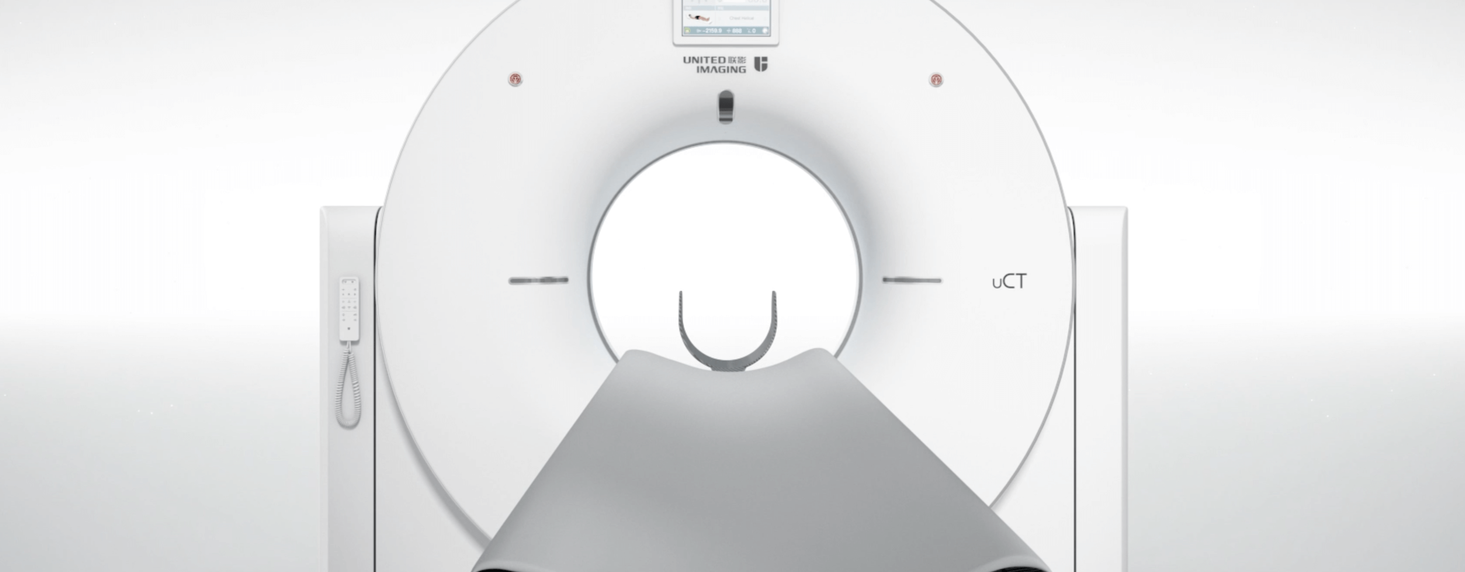
In both scans, patients lie on a moving table that slides inside the machine. During a CT scan, X-rays rotate around the patient, while MR uses strong magnetic fields and radio waves to produce images. Both CT and MR scans require the patient to remain still.
MRI and CT scans – indications and contraindications
CT is more commonly used to diagnose emergencies, while MRI is a more precise method that is ideal for diagnosing diseases of the nervous system, muscles, joints, ligaments and tendons. In terms of contraindications, MRI has limitations for patients with pacemakers or other electrical devices in the body, while CT scans are not recommended for pregnant women due to the use of X-rays.
Differences between CT and MR scan times
The duration of a CT scan is usually shorter, ranging from a few minutes to 30 minutes, while an MR scan can take from 15 minutes to up to an hour, depending on the part of the body being scanned and the type of scan. The longer duration of an MR scan is due to the need for more detailed images, which can result in slightly more discomfort for the patient.
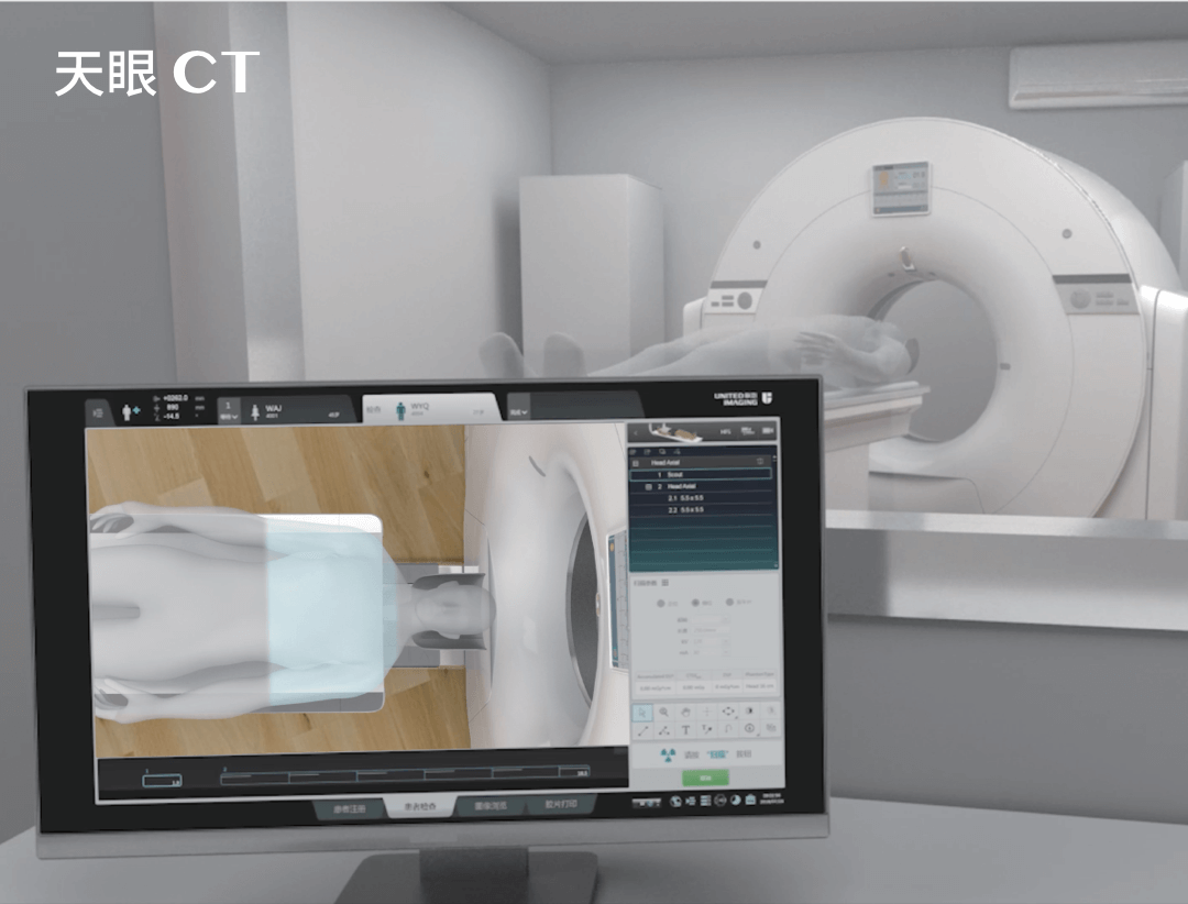
However, modern MR scanners from UIH are much faster, improving patient comfort while maintaining high image quality. In the case of CT scanners, UIH machines offer very precise results with extremely low radiation doses.
MRI versus CT scan – when are they used?
MRI is preferred for diagnosing conditions of the nervous system, muscles, joints and other soft tissues. CT scans are more commonly used in trauma, to diagnose conditions of the lungs, chest and spine, and in emergencies where a quick diagnosis is needed.
What other differences are there between a CT scan and an MRI scan?
The main difference is the imaging technique – CT uses X-rays and therefore carries a small risk related to exposure to electromagnetic radiation. Magnetic resonance uses magnetic fields and radio waves, which are considered safer, especially for patients who need frequent scans. MRI can also produce more detailed images of soft tissues, unlike CT, which is better at imaging bones and internal organs.
CT versus MRI – which scan gives more accurate results?
In general, MRI is considered more accurate for diagnosing diseases of soft tissues such as muscles, ligaments and the brain. On the other hand, CT, with its speed and ability to image bones, is better at diagnosing trauma, lung disease and other emergencies. However, the choice of method depends on the individual medical case and the doctor’s decision.
Each of these imaging modalities has its own unique advantages and limitations, making them complementary tools in medical diagnosis. The choice between them depends on the specifics of the patient’s condition, the diagnostic requirements and the patient’s overall health. At the same time, it should be noted that both CT and MRI have revolutionised medicine, enabling doctors to make more accurate diagnoses and plan more effective treatments.
*ATTENTION! The information contained in this article is for informational purposes and is not a substitute for professional medical advice. Each case should be evaluated individually by a doctor. Consult with him or her before making any health decisions.


