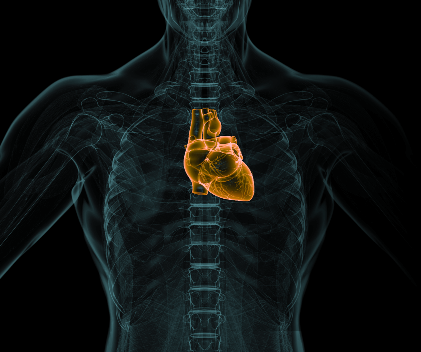
Contrast-enhanced scans. How do they work? Can anyone use contrast media?
Modern medical diagnostics very often involves imaging tests in which contrast media play an important role. These special substances are used to improve the quality of MR or CT images and allow more accurate imaging of the body’s internal structures. What exactly are contrast agents? How do they work and what are their risks and limitations?
What are contrast media and when are they needed?
In diagnostic tests, particularly computed tomography (CT) and magnetic resonance imaging (MRI), contrast agents are substances used to improve the quality of images obtained during a scan. These agents, which are usually administered intravenously, but sometimes also orally or by other means, ‘light up’ certain structures in the patient’s body, allowing for more accurate imaging and identification of potential health conditions.
Contrast agents are extremely important in the diagnosis of many conditions and diseases ranging from cancer to stroke, heart disease, haemorrhages, kidney and liver disease and more. They are particularly useful for analysing blood vessels, tumours, soft tissues and internal organs.
Which examinations use contrast agents?
Contrast media are used in many types of diagnostic tests. These include primarily:
- CT angiography – in examinations of blood vessels, contrast makes it easier to assess the patency of blood vessels and to detect stenoses, aneurysms or other abnormalities.
- Oncology – in cancer-related examinations, contrast allows a more accurate assessment of the characteristics of tumours, their vascularisation and possible metastases.
- Neurology – in examinations of the nervous system, including the brain, contrast makes it easier to detect degenerative changes and brain tumours, and to assess the condition of the brain’s blood vessels.
- Soft tissue examinations – contrast helps doctors distinguish between different structures such as muscles, tendons or internal organs, which is crucial in diagnosing abdominal and pelvic disease.
Types of contrast media
The most commonly used agents are iodine-based for CT and gadolinium-based for MRI. The choice of agent depends on the type of test and the specific area of the body to be scanned. These agents differ in their properties, such as osmolarity, which affects how they work and the potential risks to the patient.
How the contrast agent works
In CT scans, iodinated contrast agents absorb X-rays, allowing structures filled with the agent to be clearly seen. In MRI scans, gadolinium-based contrast agents affect the magnetic properties of tissues, allowing better imaging of areas of interest.
Contrast-enhanced scans – how should I prepare?
Preparation for a contrast-enhanced scan may include fasting for several hours before the scan and stopping certain medications. Each case is unique and should be discussed in detail with your treating physician.
Can anyone have a contrast scan?
Contrast-enhanced scans are generally safe and effective, but there are some contraindications that need to be considered. Although they are quickly eliminated from the body, contrast media can cause allergic reactions in some people. Symptoms of allergy can appear as early as 20 minutes after administration, so patients with a predisposition to allergy should be allergy-tested beforehand. Several other situations and conditions are also contraindicated for the administration of contrast media in imaging studies These are listed below.
Diabetes and drug interactions
Diabetics should inform their doctor of their condition as some diabetes medications, such as metformin, may interact with contrast media. In some cases, it is recommended that these medications be discontinued 48 hours before the test.
Renal function and creatinine testing
Special care should be taken in patients with impaired renal function. For this reason, a creatinine level test is required prior to a contrast-enhanced scan to assess renal function. This should be done at least 7 days before the planned MRI scan.
Hyperthyroidism
Patients with hyperthyroidism must inform their doctor of their condition before the scan. In some cases, a thyrostatic drug may need to be given to prevent the adverse effects of the iodine in the contrast agent.
Pregnancy and breast-feeding
Contrast-enhanced examinations are generally not recommended for women in the first trimester of pregnancy and for women who are breast-feeding. Due to the presence of gadolinium in some MRI contrast media, which can cross the placenta and accumulate in the foetus, contrast administration is limited to situations where it is absolutely necessary. In addition, breast-feeding women should avoid breast-feeding for 24 hours after contrast administration.
Other contraindications
Other contraindications include sickle cell anaemia, asthma, emphysema, Waldenström macroglobulinemia, seizures of cerebral origin, pheochromocytoma, glaucoma, hypertension, severe dehydration, acute and chronic circulatory failure, acute intracerebral haemorrhage, myeloma and iodine allergy. In each of these cases, the decision to administer contrast should be well considered and discussed with the doctor.
Side effects of contrast media
Although contrast media are usually well tolerated, there is a risk of side effects ranging from mild reactions such as nausea, hives, pruritus, erythema or hot flushes to more serious reactions such as bronchospasm, facial/laryngeal oedema, respiratory arrest, hypotensive shock, arrhythmia or even cardiac arrest. The European Society of Urogenital Radiology (ESUR) regularly publishes useful guidelines on the safe use of contrast media.
Importance of contrast in medical diagnosis
Contrast is a key factor in obtaining accurate images in many diagnostic tests. Contrast enhancement allows doctors to diagnose diseases more accurately and to assess the size of lesions, their location and possible metastases. It is particularly important when examining blood vessels, tumours, soft tissues and internal organs When used and monitored correctly, contrast enhancement significantly increases the efficiency and accuracy of diagnostic tests.
Safety of contrast media
Although there are some risks, contrast-enhanced examinations are considered safe when properly monitored and performed by experienced professionals. The use of contrast allows for more accurate imaging of the body’s internal structures, which contributes to better diagnosis and treatment of various conditions.
*ATTENTION! The information contained in this article is for informational purposes and is not a substitute for professional medical advice. Each case should be evaluated individually by a doctor. Consult with him or her before making any health decisions.


