CT urography – what is urinary tract tomography?
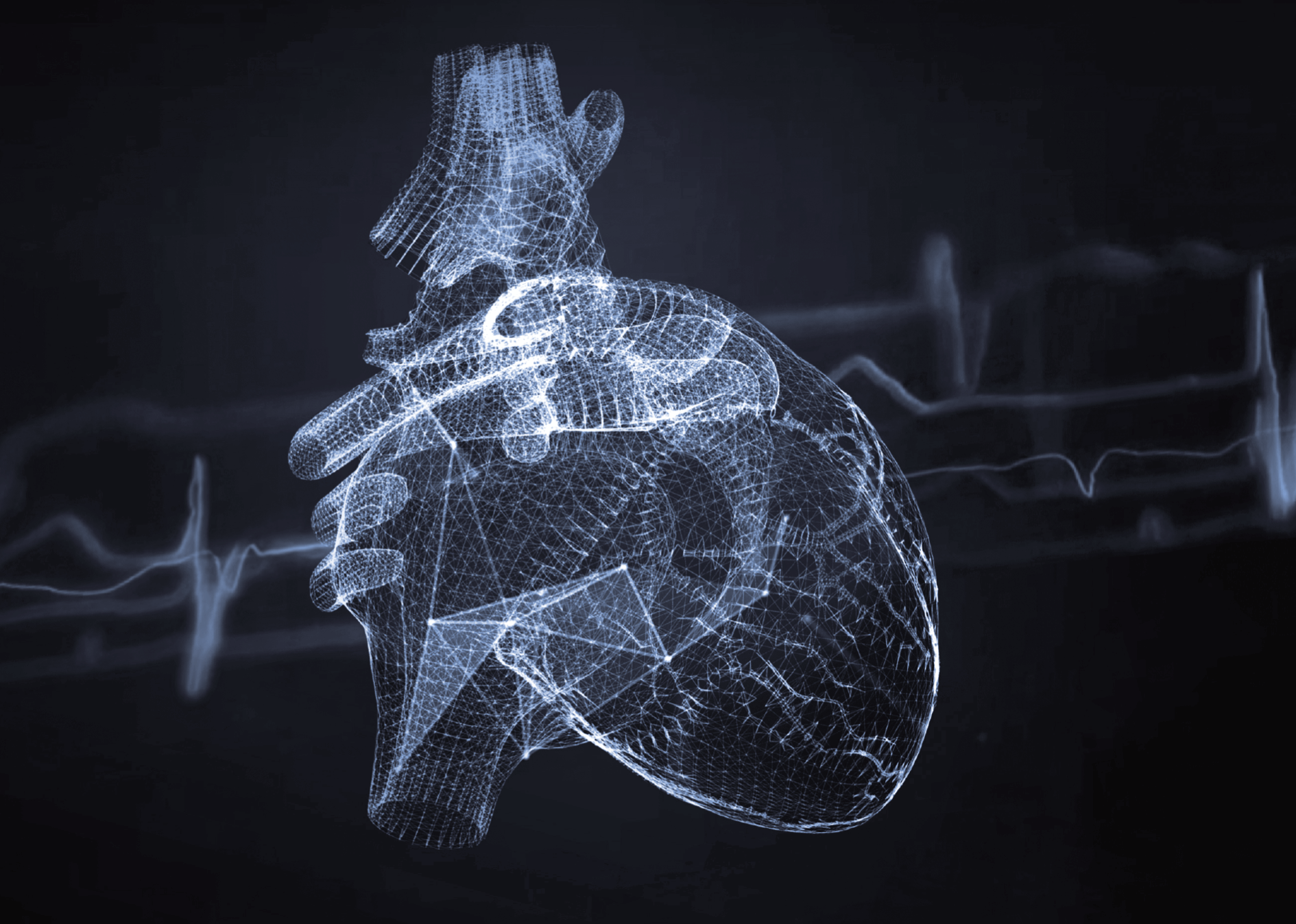
Diagnosing urinary tract disorders has become much easier thanks to advanced diagnostic techniques, one of which is CT urography. Using contrast-enhanced computed tomography, CT urography allows doctors to accurately assess the kidneys, ureters, bladder and urethra. This invaluable medical tool contributes significantly to the effective diagnosis of renal and urinary disorders and treatment planning.
What is CT urography?
CT urography is an examination that covers almost the entire urinary system, including the kidneys, ureters, bladder and part of the urethra. In cases where traditional diagnostic methods such as ultrasound or X-ray are not sufficient, CT urography provides more detailed images and allows the detection and assessment of tumours, kidney stones, urinary tract blockages and other urinary tract abnormalities.
What does CT urography consist in?
The examination begins with a series of CT scans that start above the diaphragm, cover the entire abdominal cavity and go all the way down to the bladder. CT urography uses contrast to enhance the image, allowing more accurate imaging of the kidney and urinary structures in the following four phases:
- native
- arterial
- venous
- excretory
Each of these phases provides unique information about the functioning of the urinary system.
CT urography – when is it used in diagnosis?
CT urography is an advanced diagnostic technique used in many clinical scenarios where a detailed assessment of the urinary system is required. It is mainly used when other methods, such as ultrasound or standard X-ray (digital radiography), do not provide sufficient information. CT urography is particularly useful in the diagnosis of kidney and ureteral stones, where its ability to detect even small calculi is invaluable.
It is also crucial in the detection and evaluation of kidney and bladder tumours and other pathological lesions of the urinary tract, including inflammation and infection. Doctors often rely on CT urography in cases of suspected hydronephrosis (accumulation of urine in the kidneys) and in the diagnosis of congenital anomalies of the urinary system.
Because of its high image resolution, it is also used to assess the condition of the renal blood vessels, which is important in the diagnosis of renal hypertension. CT urography is also used to plan and assess the effectiveness of surgical procedures and to monitor patients after kidney transplantation.
The ability to obtain detailed images in different phases allows a comprehensive assessment of urinary function. This imaging modality is therefore invaluable in cases where an accurate diagnosis is essential for rapid and effective treatment. It should be remembered that the decision to perform CT urography should always be preceded by a thorough analysis of the patient’s needs and condition, in order to take advantage of all the benefits of this method while minimising the risks.
How to prepare for CT urography?
Prior to the CT urography, the patient must be fasting but drinking plenty of fluids and, in the case of a contrast-enhanced examination, blood tests must be carried out. The patient must provide relevant documentation, such as a referral, results of previous tests and information about his or her medical condition. On the day of the test, the patient should avoid coffee and cigarettes and follow a light diet.
What does a CT urography scan look like?
As with any CT scan, the patient lies on a table that is automatically moved into the CT gantry. The radiology technician, who is in another room, is in constant visual and audio contact with the patient via an intercom system. If necessary, the patient may be asked to hold his or her breath for a short time to improve the quality of the images.
How long does a CT urography take?
The scan usually takes up to 30 minutes, with an exposure time of only a few tens of seconds. The patient should remain still during the scan to ensure the best image quality.
Risks and contraindications – is urography safe?
Although CT urography is generally safe, there are some risks and contraindications. The main risk is an allergic reaction to the contrast agent, especially in people who are allergic to iodine. In addition, patients with kidney failure may be at higher risk of complications. Severe liver disease, circulatory failure, hyperthyroidism and pregnancy are also major contraindications. It should also be noted that CT urography should not be repeated frequently due to the X-ray exposure involved.
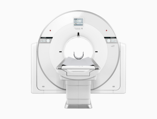
CT urography equipment
CT urography uses specialised CT scanners to produce precise images of the urinary tract. These systems are equipped with advanced data processing mechanisms and high-resolution image generation software, which allows detailed models of internal anatomical structures to be created.
In summary, CT urography is a modern, detailed and safe method of diagnosing the urinary system, allowing the condition of the kidneys, ureters, bladder and other structures to be assessed. It plays a key role in the diagnosis of kidney stones, urinary tract obstruction, infections, tumours, cysts, congenital defects and other urinary tract pathologies. The patient should always prepare properly for the examination and consult with his or her physician.
*ATTENTION! The information contained in this article is for informational purposes and is not a substitute for professional medical advice. Each case should be evaluated individually by a doctor. Consult with him or her before making any health decisions.