Abdominal CT scan
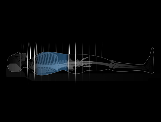
Abdominal computed tomography (abdominal CT scan) is an advanced radiological examination that allows detailed imaging of the internal structures of the abdominal cavity. CT scans, which use X-rays and computer image analysis, provide doctors with extremely precise information about the condition of organs within the abdominal cavity, including the liver, pancreas and kidneys, as well as elements of the digestive and vascular systems. They are a non-invasive and painless technology that enables quick and accurate medical diagnosis.
What can an abdominal CT scan detect?
CT scans of the abdomen can detect a wide range of conditions, including tumours (both benign and malignant), inflammations, infections, internal bleeding and blood vessel problems such as aneurysms. They are, moreover, a useful tool in diagnosing liver, pancreas, kidney and gallbladder diseases. CT scans have also been successfully used to assess patient condition after abdominal trauma, monitor the progress of treatment and plan surgical procedures.
Indications for an abdominal CT scan include:
- abdominal pain of unclear cause
- problems with the digestive system, such as bleeding, inflammatory bowel disease, Crohn’s disease or ulcers
- suspected cancer or metastases to internal organs
- assessment of trauma or internal injuries caused by an accident
- assessment of internal organs before planned surgery
- diagnostics for possible abdominal infections or inflammations
Specialised abdominal CT variants
Advanced abdominal computed tomography variants enable specialised imaging examinations of various structures and systems within the abdominal cavity to be conducted in a painless and non-invasive manner, often providing an alternative to more invasive (e.g., endoscopic) diagnostic methods.
Examples of such specialised abdominal CT examinations include:
- CT cholangiography – a CT examination of the bile ducts used to diagnose their diseases, including cholelithiasis (gallstones), tumours or cholangitis. It can also be used when planning surgical or endoscopic operations.
- CT enterography – a detailed examination of the gastrointestinal tract, especially the small intestine, together with the abdominal and pelvic organs. The scan is conducted after the patient has been administered intravenous contrast and given a solution to drink in order to fill the intestine. It is used primarily to detect inflammatory diseases of the small intestine.
- CT colonography – an examination also referred to as non-invasive virtual colonoscopy, which allows the colon to be imaged. It is used as an alternative to traditional colonoscopy in order used to detect polyps or colon cancer.
- Abdominal CT perfusion – a CT technique that measures blood flow to various organs in the abdominal cavity, which is useful in assessing blood flow in tumours or other lesions within abdominal organs. It is also used to monitor the condition of organs after transplants.
- CT urography – a state-of-the-art CT examination of the urinary system that covers the kidneys, ureters and bladder. It is performed with contrast, which allows extremely detailed images of urinary structures to be obtained in order to detect any abnormalities, such as e.g. tumours or stones.
- Abdominal angio-CT – an examination revealing the vascular anatomy within the abdominal cavity, which allows the diagnosis of the condition of blood vessels and arteries, including the detection of stenoses, aneurysms or vascular injuries. It is also used for vascular surgery planning.
- CT venography – a tomographic technique used for detailed analysis of the condition of veins, for instance to diagnose deep vein thrombosis and vascular pathologies or to evaluate veins before surgical procedures.
Will an abdominal CT scan show lesions in the intestines?
Abdominal CT scans are helpful in diagnosing various pathological conditions of the intestines, including inflammations, obstructions, perforations and the presence of tumours. When diseases and inflammatory conditions of the intestines are suspected, specialised abdominal CT examinations are primarily used to diagnose them, such as the aforementioned CT colonography for the large intestine or CT enterography for diseases of the small intestine.
How long does an abdominal CT scan take?
The duration of an abdominal CT scan depends on a number of factors, including, primarily, the scope of the scan and the need to administer a contrast agent and/or to drink a special solution that highlights the structures within the gastrointestinal tract. Usually, however, such an examination takes 15 to 30 minutes.
The scan itself is quick, but patient preparation and waiting time for the result may prolong a visit at a medical facility. For abdominal CT, it is advisable to wear loose and comfortable clothing that is free of metal parts, such as buttons or metal bra underwires, as these may interfere with examination results.
Abdominal CT scan with contrast
Abdominal CT scans can be performed both without and with a contrast agent. The decision to use contrast depends on the examination purpose, but when diagnosing tumours, bleeding or certain vascular diseases, the use of a contrast agent greatly increases the accuracy of the results obtained, which is why abdominal CT examinations are very often performed with contrast.
Is an abdominal CT scan result immediately available?
Abdominal CT results are usually not available immediately after the examination. The images need to be analysed by a radiologist, which may take several hours to several days. However, in some cases, especially emergencies, results can be obtained faster.
Which is better: abdominal CT or MRI?
Just as CT scans of the abdomen are more detailed than ultrasound, MRI enables even more detailed evaluation of tissues, making it preferable for diagnosing diseases of the liver, pancreas and vascular structures. However, it should be kept in mind that abdominal CT scans are faster, more accessible and often used in emergency situations. For examinations performed privately, CT is also cheaper, but at the same time, since computed tomography involves radiation, an abdominal CT scan requires a doctor’s referral. The final choice of diagnostic method should be made by the doctor based on an individual assessment of the patient’s condition and preliminary diagnosis.
It should be noted that United Imaging Healthcare’s modern CT scanners are able to perform in-depth analyses of abdominal structures and organs using low radiation doses. This is made possible by innovations in CT imaging technology, including the use of advanced artificial intelligence algorithms that can produce extremely accurate images without the need for high radiation doses.
*ATTENTION! The information contained in this article is for informational purposes and is not a substitute for professional medical advice. Each case should be evaluated individually by a doctor. Consult with him or her before making any health decisions.


