Head CT scan
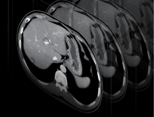
Head computed tomography (head CT) is an innovative diagnostic tool that has revolutionised the way doctors see and understand the brain. Unlike traditional X-rays, CT scans offer extremely detailed 3D images of the brain’s structures, making it possible to accurately identify and evaluate various neurological conditions.
Head CT in practice
A head CT scan is not very different from CT scans of other organs. During a head CT scan, the table on which the patient lies is slid into the CT scanner bore where he or she must remain still while the scanner, which uses X-rays, takes a series of images from different angles and in different planes.
The resulting images are then processed by sophisticated computer software to create a 3D image of the brain. This enables brain structures to be examined quickly and accurately, including areas that are difficult to access by other diagnostic methods, such as ultrasound, which cannot be used for head examinations.
Head CT applications
A head CT scan is often recommended to diagnose a variety of neurological conditions and injuries, as well as any conditions that require a detailed analysis of brain structures. Most importantly, because a computed tomography examination can be performed quickly, CT scans are particularly useful in emergencies, when an instant and accurate diagnosis is crucial to saving the patient’s life.
Brain CT scans can detect:
- brain tumours, both benign and malignant
- intracranial bleeding, including haematomas
- strokes – both those caused by haemorrhages and vascular blockages
- inflammatory conditions of the brain, for instance meningitis
- brain damage caused by trauma, such as concussions or craniocerebral injuries
- vascular anomalies, for instance aneurysms or vascular malformations
- degenerative changes and other neurological conditions, such as for instance multiple sclerosis
Indications for a head CT scan include:
- Head injuries – especially in cases of suspected serious brain damage, haematomas or skull bone fractures.
- Suspected stroke – a head CT scan can quickly identify the type of stroke (haemorrhagic or ischemic).
- Neurological symptoms such as severe headaches, seizures, changes in consciousness or other symptoms that suggest neurological problems.
- Monitoring disease progression – for patients with brain tumours or following head injuries.
- Diagnosing infections and inflammation – for instance, meningitis or encephalitis.
Specialised head CT scan variants
Computed tomography of the head performed using innovative scanners equipped with modern medical imaging software enables specialised examinations to be conducted in the brain area, such as:
- CT perfusion – a head CT scan that allows assessment of blood flow in brain tissues, which is particularly useful in diagnosing and monitoring strokes.
- CT angiography – a head CT scan that focuses on blood vessels in the brain. It can be used to detect aneurysms, vascular malformations and other vascular anomalies.
- Dynamic 4D imaging – a technology offered by UIH scanners that enables the acquisition of dynamic angiographic images for functional measurements, which is particularly useful in stroke diagnosis.
Preparing for a head CT scan with contrast
Preparation for a head CT scan is similar as in the case of other radiological examinations. For head CT scans with contrast, prior testing of creatinine levels is necessary. A doctor’s referral is required for a head CT scan.
The patient should be fasting before an examination with contrast. A head CT scan with contrast is usually used to diagnose inflammations, neoplastic lesions and possible metastases or vascular malformations. In cases of trauma or lesions in the external structures of the brain, face or skull, examinations are usually performed without administering contrast.
What does a head CT scan look like?
The examination usually takes 15–30 minutes. The patient lies down on a special table, which is slid into the CT scanner bore. Although the radiologists are in a separate room during the examination, but are in constant contact with the patient via intercom. There is no need to undress for a head CT scan, but earrings should be removed from ears, etc.
If the head CT scan is performed without contrast, no special preparations are required and the patient can leave the medical facility immediately after the examination. In the case of a head CT scan with contrast, it is advisable to stay at the medical facility for at least half an hour in case an allergic reaction to the contrast agent develops. The results of a head CT scan can usually be collected after a few days, as they require specialist interpretation and annotation.
Is a head CT scan safe?
Although a head CT scan involves exposure to X-rays, it is considered a safe examination when it is performed according to medical recommendations. Doctors consider the potential risks of radiation, taking into account the diagnostic benefits that a head CT scan offers. This examination, as well as all other imaging tests that utilise X-rays, is not indicated for pregnant women due to the risk to the developing foetus.
*ATTENTION! The information contained in this article is for informational purposes and is not a substitute for professional medical advice. Each case should be evaluated individually by a doctor. Consult with him or her before making any health decisions.


