Craniofacial computed tomography
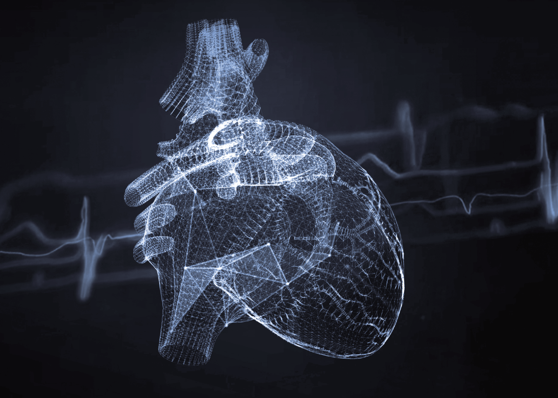
Craniofacial computed tomography is an extremely precise imaging test, which achieves a high level of detail owing to the use of multi-row CT scanners and state-of-the-art software that makes it possible to create virtual anatomical models of the complex structures of the viscerocranium: the part of the skull that surrounds our eyes, ears and the upper respiratory and gastrointestinal tracts. This examination is invaluable in diagnosing a variety of conditions, from injuries to cancerous lesions.
The complex anatomy of the craniofacial (viscerocranial) region requires a more specialised examination than a simple CT scan, and this examination is referred to as a craniofacial CT scan. This is because this part of the skull consists of multi-elemental structures that include the cartilage and bones of the face, the dental system, eye sockets, sinuses and nasal passages as well as the anterior part of the brain, including soft tissues, muscles, tendons, veins and nerves. Imaging all these structures requires equipment that can produce more precise results than a traditional CT scan.
What does a craniofacial CT scan look like?
A craniofacial CT scan involves taking a series of X-ray images from different angles, which are then digitally processed to create two- and three-dimensional models of the elements examined. Computed tomography of the craniofacial structure is therefore a much more precise examination than, for example, a classic X-ray, since this examination shows in great detail bones, sinuses and soft tissues, as well as blood vessels and nerves of the craniofacial complex. In this way, it is possible to detect abnormalities that are invisible to other methods. The examination usually consists of two sequences, with bone structures being scanned during the first sequence and soft tissues during the second one.
What can a craniofacial CT scan detect?
A craniofacial CT scan makes it possible to diagnose various types of viscerocranial injuries, inflammation, infections, tumours as well as post-traumatic lesions and abnormalities in the structure and functioning of blood vessels.
Indications for a craniofacial CT scan include:
- evaluation of craniofacial injuries
- diagnosis of facial pain or lesions of unknown origin within the face (such as swelling)
- oncological diagnosis, including localising and grading neoplastic and infiltrative lesions
- diagnosis of haemorrhages and inflammation as well as other conditions within the mouth, tongue, salivary glands, paranasal sinuses, orbits and eyeballs
- detection and evaluation of congenital defects
- preparation for surgical procedures
- location of foreign bodies, for instance, in the orbit, nose or sinuses
- treatment monitoring
Craniofacial CT scan vs. head CT scan
A craniofacial CT scan differs from a head CT scan both in its scope and in imaging detail. A craniofacial CT scan focuses on the precise evaluation of the bony and soft structures of the face and skull, accounting for the complexity of its structure, which includes numerous cavities, arches and folds. A head CT scan, on the other hand, is a more general examination that focuses on the entire head, including the brain and other deeper structures. A craniofacial CT scan uses different parameters than a whole head CT scan, which enables even very small lesions to be detected.
Preparation for craniofacial computed tomography with and without contrast
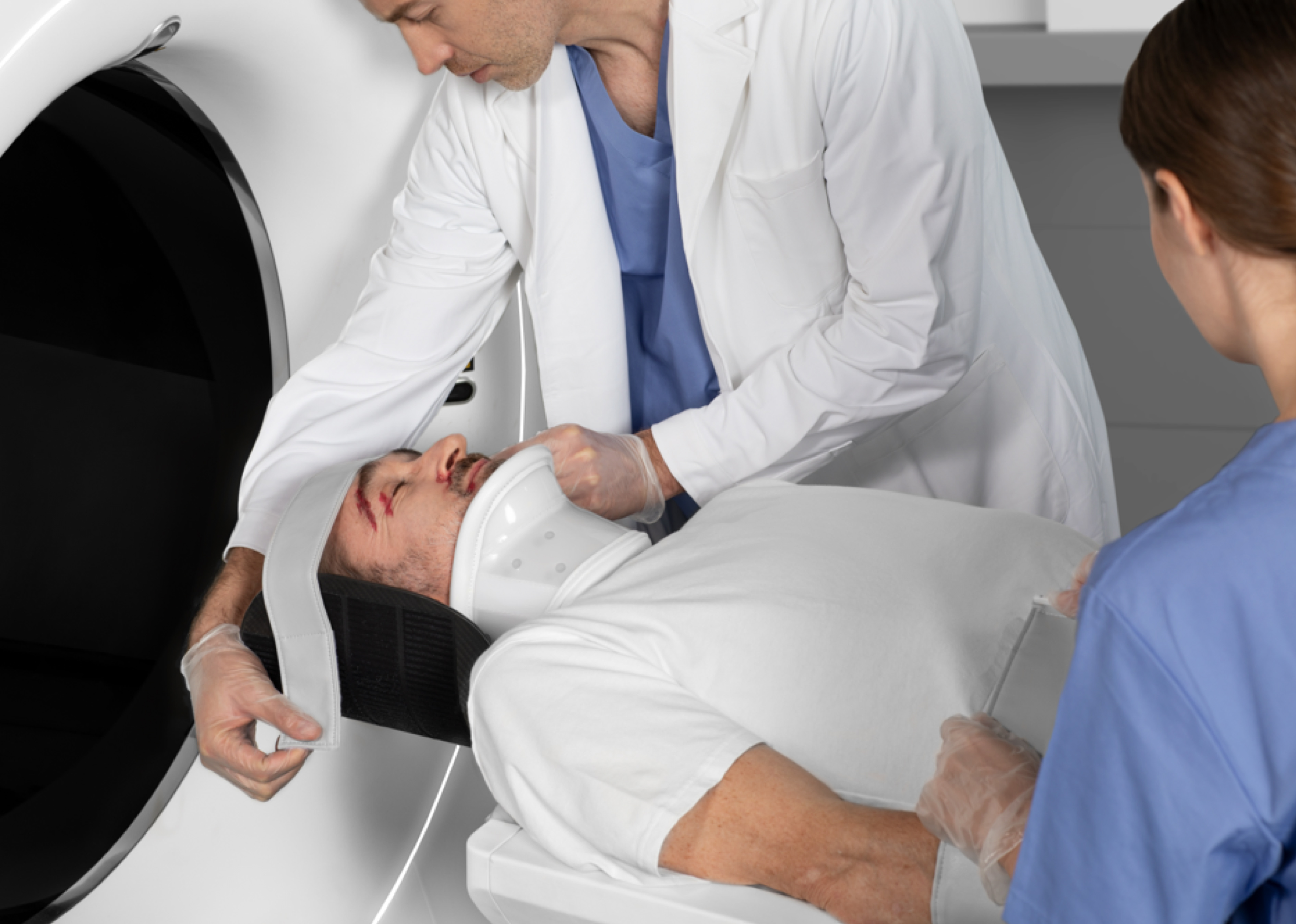
Depending on the purpose of the examination, a craniofacial CT scan can be conducted with intravenously administered contrast, which improves the visibility of internal structures, or without contrast. The preparation for a craniofacial CT with contrast includes being fasted and undergoing tests to confirm that the kidneys are functioning efficiently enough to filter the contrast agent out of the blood. The patient should also remove any metal objects from the head region, including not only jewellery, but also eyeglasses or metal braces.
The administration of a craniofacial CT contrast agent is recommended when diagnosing cancers, infections, inflammations and vascular abnormalities. In post-traumatic cases and those involving the evaluation of bone and soft tissues, contrast is usually not required.
A craniofacial CT scan – what does it look like? Is referral required? How long does it take to get results?
A craniofacial CT scan is a radiological examination, and thus it requires a doctor’s referral. The use of United Imaging Healthcare’s modern CT scanners significantly reduces the radiation dose during this type of examination, making it safer and faster, but X-rays are emitted nevertheless.
The examination itself takes from 15 to 30 minutes and looks similar to a standard head CT scan, but scanner settings are different. As a rule, it takes several days for the results to be available: although the images are produced immediately, they must be annotated by a radiologist.
Craniofacial CT scan – contraindications
Contraindications include pregnancy, allergy to iodine, hyperthyroidism, kidney failure and the presence of metal implants. In some cases, because of the risks, doctors may recommend alternative diagnostic methods. An alternative to a craniofacial CT scan is MRI, which provides even better diagnostic results.
*ATTENTION! The information contained in this article is for informational purposes and is not a substitute for professional medical advice. Each case should be evaluated individually by a doctor. Consult with him or her before making any health decisions.