Computed tomography of the heart
A cardiac CT scan is a specialised, non-invasive diagnostic imaging test that uses X-rays to create detailed images of the heart. It is particularly useful in diagnosing coronary artery disease and assessing the risk of myocardial infarction, as well as in diagnosing heart defects and evaluating the patient’s condition after cardiac surgery.
What does a cardiac CT scan look like?
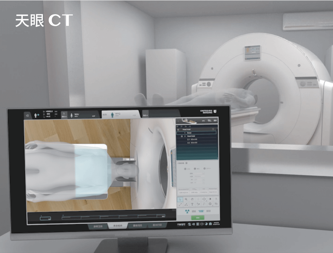
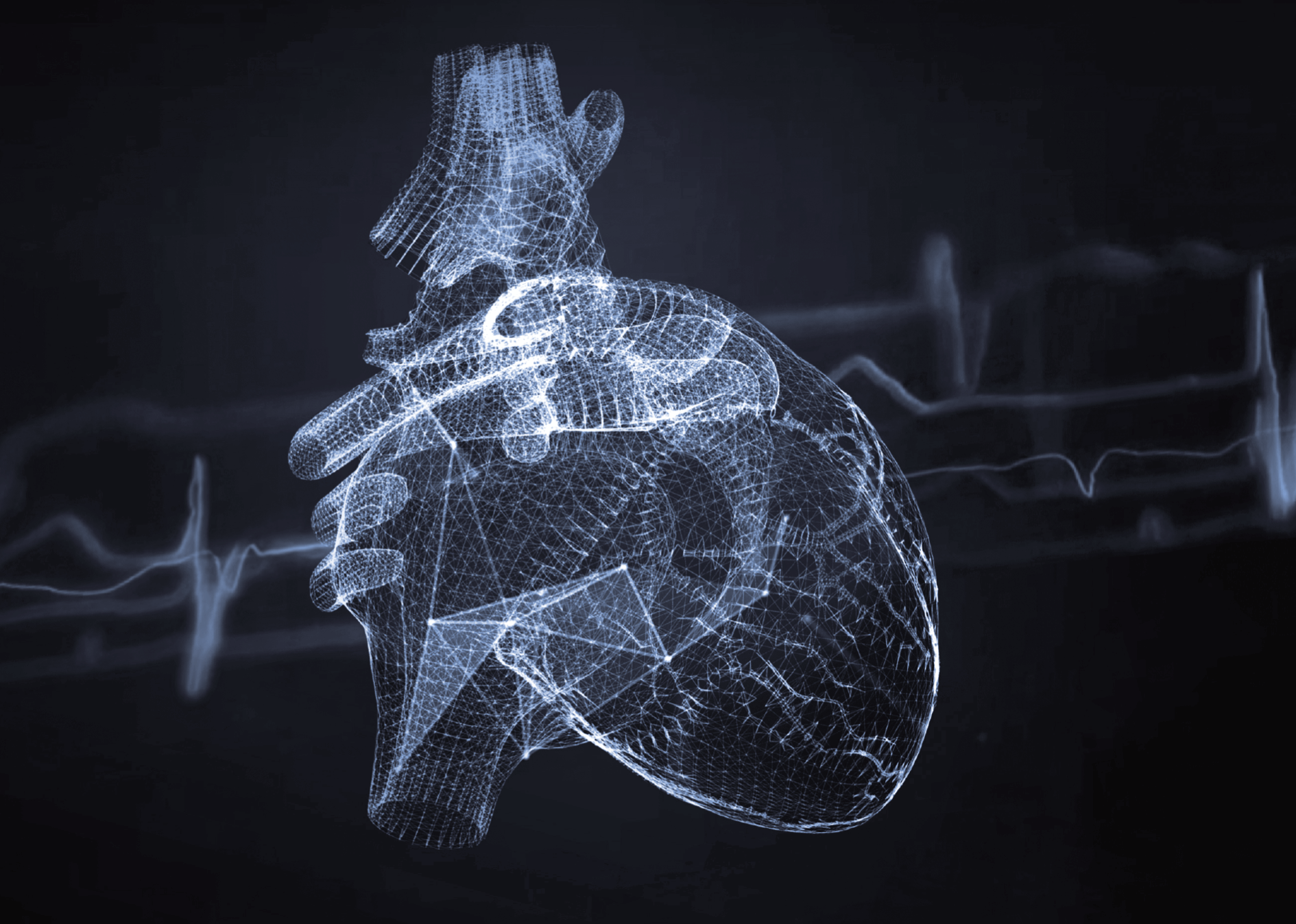
Computed tomography of the heart is an advanced medical imaging technique that enables the structure of the heart and surrounding blood vessels to be visualised in detail. The procedure uses X-rays and computer technology, including state-of-the-art software, to produce detailed cross-sectional images of the heart, which enable in-depth quantitative and functional analysis.
Cardiac CT is particularly useful in assessing the condition of the coronary vessels, looking for stenosis or blockages, as well as in evaluating other heart structures such as the ventricles, valves or walls. A cardiac CT scan enables the detection and evaluation of a variety of cardiac pathologies.
Indications for a cardiac CT scan include:
- Assessing the degree of coronary stenosis and diagnosing coronary artery disease.
- Detecting calcification in coronary arteries, which may indicate the risk of cardiovascular disease.
- Evaluation of structural anomalies of the heart, including congenital defects.
- Diagnosing heart tumours.
- Evaluating the function and structure of heart valves.
- Evaluation of systolic and diastolic parameters
- Monitoring the progress of heart disease treatment, including by-pass monitoring.
With its high resolution and 3D imaging capabilities, a cardiac CT scan provides valuable information that can be crucial in making therapeutic decisions. Cardiac CT can be used to diagnose atherosclerosis, abnormal valve function and pericardial disease as well as aortic aneurysms, post-infarction scarring or pulmonary embolisms.
Indications for a cardiac CT scan include:
- coronary heart disease risk
- situations where other tests do not provide a clear diagnosis with symptoms such as dyspnoea (shortness of breath), rapid fatigue and breathlessness or chest pains
- monitoring after bypass surgery
- assessment of cardiac function
- evaluation before cardiac surgery
- suspected coronary artery anomalies
Computed tomography of the heart versus chest CT scan
Cardiac CT and chest CT differ in their examination scopes and specific imaging parameters. A cardiac CT scan focuses on accurate imaging of the heart and coronary vessels, enabling the assessment of the structure of the heart, the condition of the blood vessels, and the presence of calcium deposits. A chest CT scan, on the other hand, covers a wider area, including the lungs, bronchi, aorta, thoracic bones and other structures in this body region. Therefore, it is mainly used to diagnose pulmonary conditions and other chest pathologies.
Thus, despite some similarities, these two examinations differ in their diagnostic goals and in the detail with which specific structures are imaged. In addition, during a cardiac CT, an ECG is also usually performed, meaning that electrocardiograph electrodes are attached to the patient’s chest, and blood pressure and saturation are measured as well.
Cardiac CT vs. coronary angiography
A cardiac CT scan is a non-invasive diagnostic method that uses X-rays to create detailed images of the heart and blood vessels. It is particularly useful in assessing the structure and condition of coronary arteries, as well as in diagnosing cardiovascular disease. A major advantage of a cardiac CT scan is its ability to quickly and accurately detect calcifications in the coronary arteries, which may provide an early indicator of atherosclerotic coronary artery disease.
A cardiac CT scan is less invasive than coronary angiography and the recovery time required is usually shorter. During a coronary angiography, which is an invasive diagnostic procedure that also uses contrast and X-rays to visualise the inside of the coronary arteries, a contrast agent is injected directly into the coronary arteries through a catheter, which is associated with a higher risk of complications after the examination.
Cardiac CT scan – with or without contrast?
Cardiac CT with contrast is usually used when a thorough evaluation of the heart’s blood vessels is required, for instance to detect stenosis or blockages in the coronary arteries. This type of examination enables the assessment of blood flow in the heart and detection of ischaemic heart disease.
Cardiac CT without contrast, on the other hand, is mainly used to evaluate the structure of the heart, for instance to assess calcification in the coronary arteries, which can be an indicator of cardiovascular disease risk. This examination is safer and is possible also in cases where the use of a contrast agent is contraindicated.
Contraindications to a cardiac CT scan:
- pregnancy (due to the risk of foetal exposure to radiation)
- kidney failure, as the contrast agent used in CT can affect kidney function
- hypersensitivity to the contrast agent in cases of allergy to iodine-containing contrast agents
- recent history of myocardial infarction/acute coronary syndrome
- cardiac arrhythmias (e.g., overly elevated heart rate or atrial fibrillation), since these can affect the quality of imaging
- severe systemic conditions that may increase the risk of complications
A doctor’s referral is required for a cardiac CT scan.
*ATTENTION! The information contained in this article is for informational purposes and is not a substitute for professional medical advice. Each case should be evaluated individually by a doctor. Consult with him or her before making any health decisions.