Breast MRI – indications, what does it look like, how to prepare for the scan and how long does a MRI scan take?
Magnetic resonance imaging (MRI) is a key medical diagnostic tool enabling detailed evaluation of breast tissues and structures. Thanks to its high resolution and ability to acquire images in different planes, breast MRI enables lesions to be accurately located, tumours to be identified and their characteristics to be evaluated as well as treatment response to be monitored. This is especially important for detecting breast cancer in early stages, which in turn can increase the effectiveness of treatment and improve patient outcomes.
Breast MRI can also be useful in monitoring breast lesions in patients with a genetic predisposition to breast cancer and in evaluating breast implants. Thanks to its non-invasive nature and the fact that no ionising radiation is involved, the examination is a safe and effective diagnostic method, which can help doctors detect, monitor and treat breast diseases.
Breast MRI indications
Breast MRI, also known as MR mammography, has clear clinical indications. This examination is used to stage certain types of breast cancer and exclude the presence of multiple tumour foci, as well as to differentiate recurrent cancer from scarring in women after breast surgery.
Another indication for an MRI scan is a negative mammogram and ultrasound where the presence of a metastatic lymph node has been confirmed. Breast MRI is also used for the following purposes:
- monitoring early response to chemotherapy;
- as a screening method for women at increased risk of breast cancer, particularly those with genetic mutations associated with a higher incidence of breast cancer, such as BRCA-1 and BRCA-2 mutations;
- assessing the condition of breasts reconstructed after mastectomy.
Contraindications to MRI scans
The scan is not performed if factors are found that may increase the risk of complications or impede the examination. These include:
- the presence of a pacemaker, artificial valves, clips or vascular stents;
- the presence of a neurostimulator or insulin pump;
- the presence of a cochlear implant;
- the presence of screws, endoprostheses, stabilisers, implants or certain types of dental fillings;
- the presence of an intrauterine contraceptive device.
Other contraindications include the presence of metal debris in the body, intolerance to contrast agents or claustrophobia, which can cause discomfort or anxiety while the patient lies inside the narrow bore of an MRI machine.
Menstruation is not an absolute contraindication to MRI, but it may affect the patient’s comfort, and therefore, many medical facilities recommend avoiding the examination during menstruation if possible.
READ MORE: CAN AN MRI BE PERFORMED DURING PREGNANCY?
How to prepare for a scan?
Several days before the scheduled examination, kidney function must be assessed. The patient is referred for a blood test so that her creatinine levels can be measured. In addition, in some cases the glomerular filtration rate (GFR) should also be determined, which provides information about the kidneys’ ability to remove toxins from the body. These tests are important because some of the contrast agents used during an MRI scan are excreted with urine and this puts additional stress on the kidneys. It is therefore crucial to make sure that the patient’s kidneys are functioning properly in order to avoid possible complications and ensure the safety of the diagnostic procedure.
The patient should appear for the examination fasted, in loose clothing that does not include metal parts, i.e. buttons, zippers or jewellery. On the day of the examination, cosmetics that contain metallic substances should also be avoided to prevent interference with the images obtained. If the patient has undergone previous surgery, especially in the breast area, the medical staff should be informed in order to ensure safety and proper interpretation of the images obtained.
MORE ON THIS SUBJECT: HOW TO PREPARE FOR AN MRI SCAN?
What does a breast MRI look like and how long does it take?
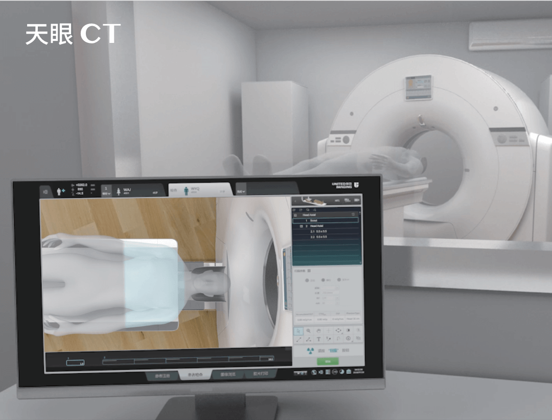
During the scan, the patient is placed on the examination table, which moves inside a cylindrical MRI machine bore. The examination usually lasts between 20 and 60 minutes, but its duration may vary depending on the diagnostic needs and the complexity of the diagnostic procedure. During this time, the patient must remain motionless, in a stable position. Different imaging sequences can be used during the examination, allowing detailed information on breast tissues and structures to be obtained.
The examination is completely painless, but the MRI machine generates loud and quite unpleasant sounds that can cause discomfort. This discomfort can be reduced by wearing earplugs or headphones used for communication between the patient and the radiologist who is supervising the examination from an adjacent room.
If any concerning symptoms accompany the administration of contrast, the patient should immediately inform the medical staff. The patient should also increase her fluid intake in the days following the examination, which will help flush the contrast agent out of her system faster. Breastfeeding mothers should also stop feeding for the next two days, and the milk drawn during this time should be disposed of.
Breast MRI results are usually available a few days after the examination. However, the waiting time may vary depending on the specific circumstances and the medical facility where the examination was performed. Some facilities may be able to provide results more quickly, especially if the scan was performed due to urgent diagnostic needs.
*ATTENTION! The information contained in this article is for informational purposes and is not a substitute for professional medical advice. Each case should be evaluated individually by a doctor. Consult with him or her before making any health decisions.
THIS MAY ALSO INTEREST YOU: ABDOMINAL MRI SCAN


