What are the principles underlying magnetic resonance diffusion-weighted imaging?
Diffusion-weighted imaging (DWI) is an innovative technique used in magnetic resonance imaging (MRI). It allows for an extremely accurate assessment of the movement of water molecules in tissues, which is crucial in diagnosing various brain disorders, but other conditions as well. If you want to learn what this examination consists of and when it is most often used, read on!
What is diffusion-weighted imaging?
Diffusion-weighted imaging is a specialist magnetic resonance (MR) technique that enables the movement of water molecules in tissues to be assessed. As a result, information can be obtained about tissue structure and function at the molecular level. In medical practice, diffusion-weighted imaging has become an indispensable tool in the diagnosis of many conditions, especially those of the nervous system and brain, but other ones as well. Before we get into the details, it is worth clarifying a few points about what exactly this type of examination involves.
How does MRI with diffusion-weighted imaging work?
During a magnetic resonance examination with diffusion-weighted imaging, the patient is placed in an MRI machine that generates a magnetic field. During the examination, the machine processes signals from water molecules in the patient’s body. The diffusion-weighted imaging (DWI) sequence uses magnetic gradients to detect the movement of water molecules. This process results in images that show areas with varying degrees of diffusion in the tissues.
MR diffusion versus MR perfusion
Although the terms ‘diffusion’ and ‘perfusion’ are sometimes used interchangeably, they actually refer to two different phenomena and examinations. This is because diffusion is the free movement of water molecules in tissues, while perfusion is the flow of blood through blood vessels. In the context of magnetic resonance imaging, diffusion-weighted imaging (DWI) focuses on particle motion, while perfusion-weighted imaging (PWI) is typically used to evaluate blood flow.
DWI sequence in MRI and its application
Diffusion-weighted imaging is a state-of-the-art sequence with tremendous diagnostic potential that can significantly improve the accuracy of MR examinations. DWI makes it possible to detect early ischemic lesions in the brain, which is particularly useful in diagnosing stroke. In addition, diffusion-weighted imaging enables a hemorrhagic stroke to be distinguished from an ischemic stroke. In clinical practice, DWI is also used to detect tumours, abscesses and other pathological changes in various organs, such as the liver and kidneys.
Diffusion-weighted imaging in brain examinations
The brain is one of the organs that is most commonly examined using the DWI technique. Since it is highly sensitive in detecting early ischemic changes, diffusion-weighted imaging has become the standard tool for diagnosing early stroke. In addition, diffusion-weighted imaging makes it possible to assess the integrity of the brain’s white matter, which is crucial when examining patients with neurodegenerative diseases.
Advantages of diffusion-weighted imaging:
- Early stroke detection – DWI is particularly sensitive to very early ischemic lesions in the brain, allowing rapid diagnosis of stroke.
- Differentiating between types of stroke – this technique can detect hemorrhagic stroke and distinguish it from ischemic stroke.
- Detecting pathological changes – DWI can be used to detect tumors, abscesses and other lesions in various organs.
- Non-invasiveness – it is a non-invasive technique, meaning that it does not require the introduction of any foreign bodies into the patient’s body.
- High image resolution – the DWI technique allows high-resolution images to be obtained for more accurate diagnosis.
It should also be mentioned that diffusion-weighted imaging is usually combined with other MRI sequences, which allows extremely precise results to be obtained. DWI is very often used alongside the STIR sequence (a technique used to examine soft tissues) and with the already mentioned perfusion imaging. In the latter case, diffusion and perfusion can be combined in the preoperative diagnosis of intracranial tumours, providing more specific and accurate examination results than conventional MRI.
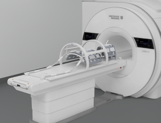
What MRI machines are equipped with diffusion-weighted imaging functionality?
Most modern MRI machines are equipped with diffusion-weighted imaging functionality. However, not all of them offer the same quality of DWI images. Today’s higher-powered MRI machines (e.g., 2 T and 3 T ones) offer better image resolution and shorter examination times compared to older models. Other things worth checking before proceeding with diffusion-weighted imaging include MRI machine specifications and available examination protocols.
Today’s diffusion-weighted imaging technologies and methods enable doctors to make more accurate diagnoses and plan treatment better for patients with various conditions. Owing to continued advances in MRI, diffusion-weighted imaging is becoming increasingly precise and accessible to a wide range of patients.
If you are looking for more information about MRI machines that enable precision diffusion-weighted imaging, feel free to contact the experts at United Imaging Healthcare who will be able to answer all your questions.
*ATTENTION! The information contained in this article is for informational purposes and is not a substitute for professional medical advice. Each case should be evaluated individually by a doctor. Consult with him or her before making any health decisions.