X-ray of the upper gastrointestinal tract
X-ray examination of the oesophagus is one of the basic tests used in diagnosing the upper gastrointestinal tract. A specialist’s referral is required for this examination, irrespective of whether it will be funded by the National Health Service or paid privately.
X-ray of the upper gastrointestinal tract is performed in the diagnosis of severe and chronic diseases of the digestive system. It involves a series of images of the upper gastrointestinal tract taken after oral administration of a contrast agent, which is usually barium sulphate suspension. This examination makes it possible to visualise the shape of the oesophagus, stomach and duodenum, and to assess their peristalsis and possible strictures.
An X-ray of the upper gastrointestinal tract includes the oesophagus, stomach and duodenum.
Oesophagus – what is it?
The oesophagus is a segment of the digestive tract that acts as a link between the pharynx and the stomach. It consists of two layers of muscular membrane that allow food movement, and an inner mucous membrane that lines its interior.
The main function of the oesophagus is to transport food and fluids from the pharynx to the stomach through coordinated peristaltic movements. This process involves rhythmic contractions of muscles that move food along the oesophagus and then into the stomach. The oesophageal wall does not absorb or digest food, and its main task is to efficiently transfer it to the next stage of digestion.
Diagnosis and diseases of the oesophagus
Diseases of the oesophagus can include a variety of problems, such as structural abnormalities (e.g., strictures or diverticula), functional abnormalities (e.g., oesophageal muscle dysfunction) or damage to individual parts (e.g. inflammation or ulcers). Symptoms of these conditions may include difficulty swallowing, pain, heartburn and other problems related to moving food through the digestive tract.
Some of the most common oesophageal conditions include gastroesophageal reflux disease, oesophageal candidiasis, oesophageal varices and oesophageal achalasia, among others.
Endoscopic examination plays a key role in the diagnosis of many oesophageal conditions. However, X-ray imaging studies are also used when certain diseases are suspected.
Indications for oesophageal X-ray examination
Today, upper gastrointestinal contrast studies are much less frequently required than in the past due to technological advances and the availability of modern diagnostic methods. Modern endoscopy, which allows direct visualisation of the inside of the gastrointestinal tract, has become the tool of choice in diagnosing diseases of this tract, reducing the need for contrast studies.
Contrast examination, which involves the use of a contrast agent to obtain a better image of gastrointestinal tract structures, is only performed in certain situations. First of all, it is used for patients in whom endoscopy is not possible for various reasons, such as technical problems, anatomical difficulties or health contraindications.
In addition, a contrast study may be necessary when endoscopy has been unsuccessful, for instance when the images obtained are insufficient to make an unequivocal diagnosis. In such cases, a contrast study may supply additional information that helps assess the patient’s condition more accurately.
Contraindications for oesophageal X-ray
Pregnancy is a contraindication to a gastrointestinal contrast study. Therefore, in menstruating women, such an examination should only be performed in the first 10 days of the menstrual cycle or after a pregnancy test has confirmed that the patient is not pregnant (unless there is a life-threatening condition).
Complete obstruction of the gastrointestinal tract is a contraindication to the use of oral contrast agents, which in turn precludes the performance of contrast studies of the upper gastrointestinal tract and of the small intestine.
How to prepare for the examination?
The patient must be fasting before the examination, which means that on the day of the examination he or she must not eat, drink or smoke cigarettes; if possible, the patient should also avoid taking medication. The day before the examination, the patient should also avoid eating foods that may lead to excessive gas production, such as carbonated beverages, fruits, dairy products or fresh bread. On the day before the examination, it is recommended that the patient take preparations that reduce bloating and gas accumulation in the intestines, such as simeticone.
What does an X-ray examination of the oesophagus look like?
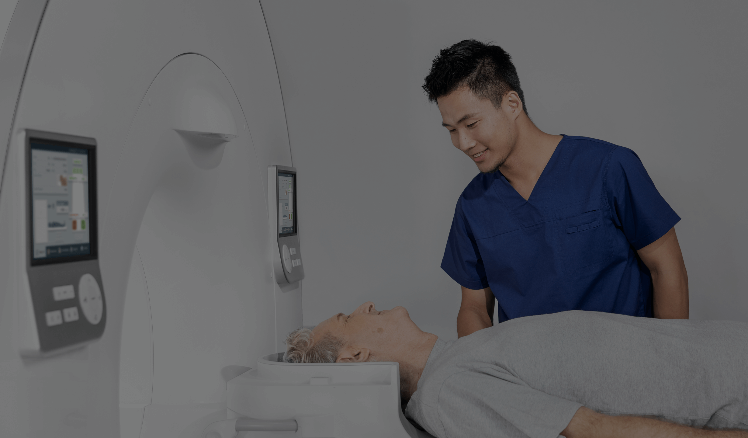
The examination is painless and only requires that the patient remain motionless in a standing or sitting position and follow the instructions of the radiologist or electroradiologist. When instructed, at the appropriate time, the patient should swallow the contrast agent in the form of a thick liquid and hold his or her breath. In modern digital X-ray systems, the imaged body parts are visible on a computer monitor immediately after the X-ray has been taken.
*IMPORTANT! The information contained in this article is for informational purposes only and is not a substitute for professional medical advice. Each case should be evaluated individually by a doctor. Consult with your doctor before making any health decisions.


