A PET/CT scan – what can it reveal, what are the indications and contraindications, and how should I prepare for it?
A PET/CT scan, which combines positron emission tomography (PET) and computed tomography (CT), is a breakthrough in diagnostic imaging that allows the precise localisation and assessment of tumours, neurological conditions and heart disease. This innovative examination, which combines the benefits of both technologies, allows both the anatomical structure and metabolic activity of tissues to be visualised, which is invaluable for planning treatment and monitoring its effectiveness. What exactly does the PET/CT scan reveal? What are its indications and contraindications? And how should I prepare for it to make sure that the diagnosis is as reliable as possible?
Positron emission tomography combined with CT
Positron emission tomography combined with computed tomography (PET/CT) is an innovative diagnostic method that can accurately determine the location and progression of cancer. It uses molecular imaging to analyse cell metabolism, combined with the accurate anatomical imaging provided by tomography. This makes it possible to assess how the disease is affecting bodily function and where exactly the pathological lesions are located.
During the PET/CT scan, a radiopharmaceutical is administered to the patient, which is taken up by cancer cells due to their increased metabolism. A CT scan performed at the same time provides anatomical images that are then combined with the PET results to make an accurate diagnosis. PET/CT is therefore particularly useful in oncology, but is also used in neurology and cardiology.
Indications for PET/CT scans – what can it reveal?
PET/CT is mainly used in oncological diagnostics because it allows extremely early detection of cancerous lesions, assessment of disease progression, precise treatment planning and monitoring of treatment effectiveness. In oncology, PET/CT is invaluable in identifying primary sites, assessing possible relapse and detecting metastasis or recurrence of the cancer.
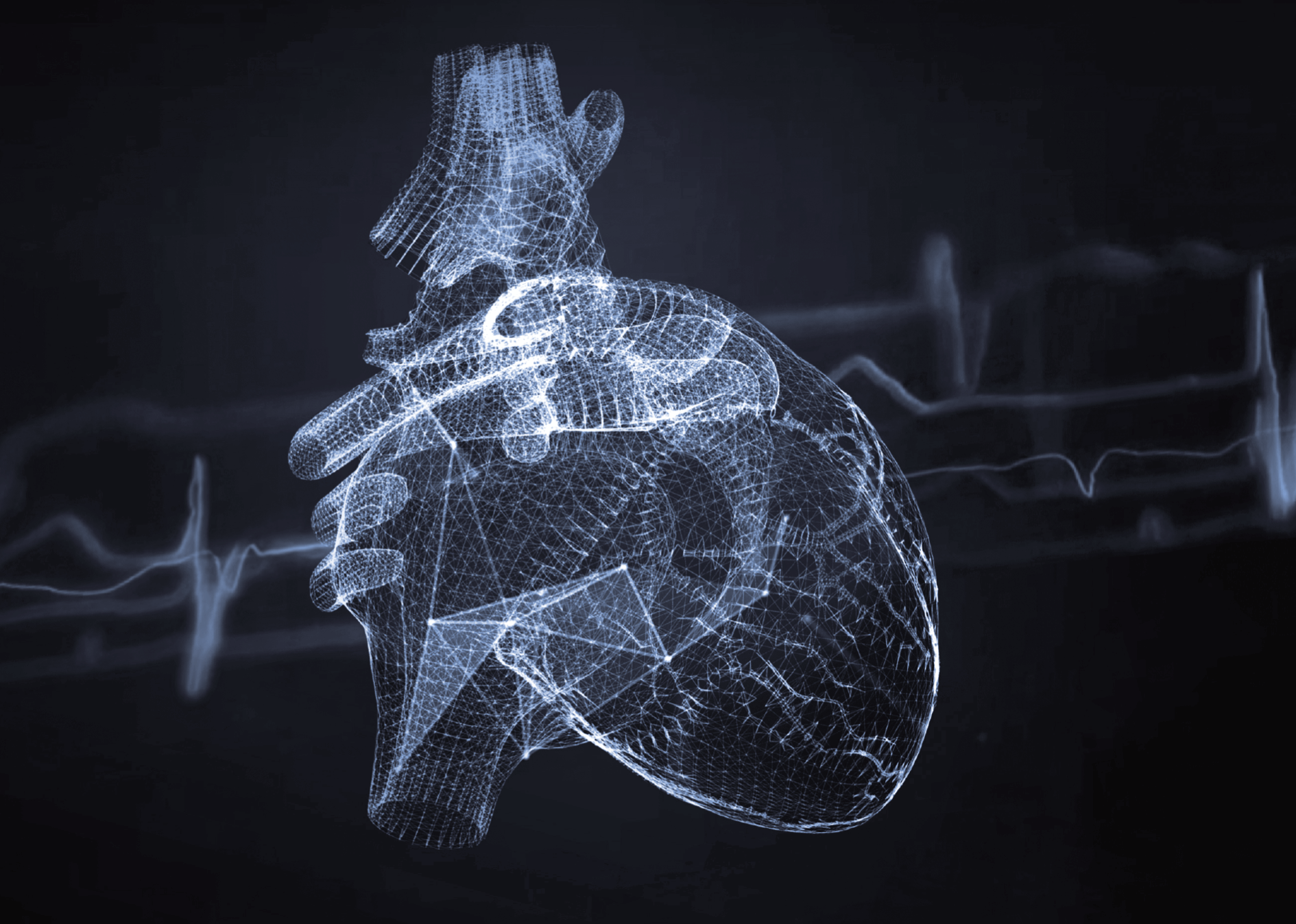
Outside of oncology, PET/CT can also be used to diagnose neurodegenerative diseases such as Alzheimer’s and Parkinson’s, as well as various forms of schizophrenia and epilepsy. In cardiology, PET/CT can be used to assess myocardial viability and diagnose cardiovascular disease.
How should I prepare for a PET/CT scan?
Preparation for a PET/CT scan includes a low-carbohydrate diet, which should be followed for 24 hours before the examination. The patient should refrain from eating and chewing gum for 6 hours before the scan, and should drink about 1.5 litres of non-carbonated mineral water. Patients taking medication should take it at normal times and only with water. It is also important to avoid strenuous exercise for 24 hours before the scan.
Diabetics should inform the medical staff of the medication they are taking and their blood sugar levels. On the day of the scan, it is advisable to eat a light breakfast (if the scan is to be done later in the day) and to take standard diabetes medication. The patient should not eat anything or take insulin for 4 hours before the scan.
What does a PET/CT scan look like?
The PET/CT scan begins with the administration of a radiopharmaceutical, after which the patient rests for about an hour to allow the substance to be distributed throughout the body. The patient then lies down on the PET/CT scanner bed, usually on his or her back, and must remain still for about 20 minutes. During the scan, a technician, a doctor and a nurse watch over the patient, and all instructions are given by intercom.
After the test, the patient can go home and resume normal activities and diet. It is recommended that the patient stays away from children, adolescents and pregnant women for 12 hours after the scan and drinks plenty of fluids so that the radiopharmaceutical is excreted quickly in the urine.
Contraindications to PET/CT
There are no absolute contraindications for CT or PET/CT examination.
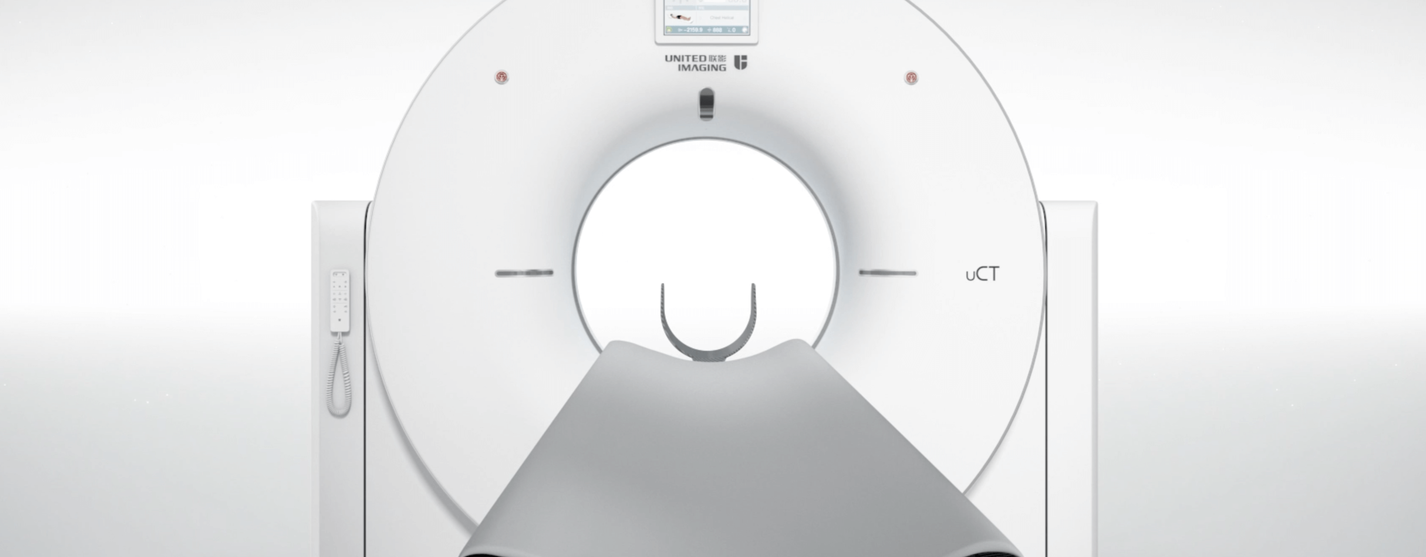
PET/CT machines
Today’s PET/CT machines are advanced hybrid scanners that combine PET and CT technology. These scanners can produce detailed anatomical images of the body and provide information about the metabolic processes taking place in its cells. The use of advanced PET/CT scanners is essential for the accurate diagnosis and monitoring of various diseases, especially cancer.
United Imaging Healthcare offers innovative PET/CT scanners that provide thorough and accurate examinations with minimal radiation exposure. Have more questions? Do not hesitate to contact us!
*ATTENTION! The information contained in this article is for informational purposes and is not a substitute for professional medical advice. Each case should be evaluated individually by a doctor. Consult with him or her before making any health decisions.