Computed tomography of the elbow
Computed tomography (CT) of the elbow joint is an advanced, rapid and non-invasive diagnostic test that enables elbow bones and joints to be imaged in detail. This examination uses X-rays and a computer to create cross-sectional images that can be transformed into a three-dimensional model of the elbow joint.
What are the indications for a CT scan of the elbow?
Computed tomography of the elbow joint is indicated in cases where other imaging modalities, such as standard X-rays or ultrasound, do not provide sufficient information. This modality can be used for diagnosing:
- bone fractures when a standard X-ray does not accurately show the fracture line or its direction;
- degenerative changes in order to assess the progression of osteoarthritis;
- post-traumatic lesions in order to evaluate damage resulting from sports injuries or accidents;
- joint and tendon injuries: CT can show details of damage to soft tissues, although magnetic resonance imaging (MRI) is more commonly used for this purpose;
- pre-operative evaluation in order to thoroughly understand the anatomy and extent of damage prior to surgery.
Computed tomography involves exposure to X-rays, which is higher than in the case of a standard X-ray, but still at a level considered safe in medicine. People with allergies to contrast media may experience an allergic reaction, so the patient should always inform the physician about such allergies before the examination.
Contraindications to CT scans
Computed tomography (CT) is a commonly used diagnostic test, but there are some contraindications and limitations that should be considered before performing a CT scan. Contraindications can be absolute (completely preventing the examination from being carried out) or relative (requiring special caution or additional evaluation).
Absolute contraindications
Because of the risk of foetal exposure to X-rays, CT is contraindicated in pregnant women unless the benefits of the examination far outweigh the potential risks. When imaging is necessary in pregnancy, magnetic resonance imaging (MRI), which does not use radiation, is often preferred.
Relative contraindications
- Contrast allergy – people with an allergy to iodine, which is present in most contrast media used in CT, are at risk of having an allergic reaction. In such cases, the physician may decide to use premedication (antihistamines and corticosteroids) or opt for an alternative imaging modality.
- Renal failure – administration of contrast may impair renal function, especially in patients with existing renal failure. Before performing contrast-enhanced CT in such patients, it is necessary to check their renal function (assessing creatinine levels and eGFR values). In some cases, the examination can be performed without contrast or using other contrast agents.
- Diabetes – diabetic patients who are using metformin should inform their physician about this fact. In rare cases, contrast administration can lead to lactic acidosis, and thus it may be necessary to discontinue metformin for a short time before and after the examination.
- Thyroid diseases – in patients with hyperthyroidism, contrast administration can trigger a thyroid storm, which is a life-threatening condition. In such patients, caution and prior preparation are necessary.
What does an elbow CT scan look like?
Prior to the examination, the patient may be asked to provide the results of previous imaging studies (such as X-rays) and other relevant medical documents. Subsequently, the technician or physician conducts a brief interview, asking about contraindications. The patient is asked to remove any metal objects (jewellery, watch) from the examined area, as they may interfere with the image.
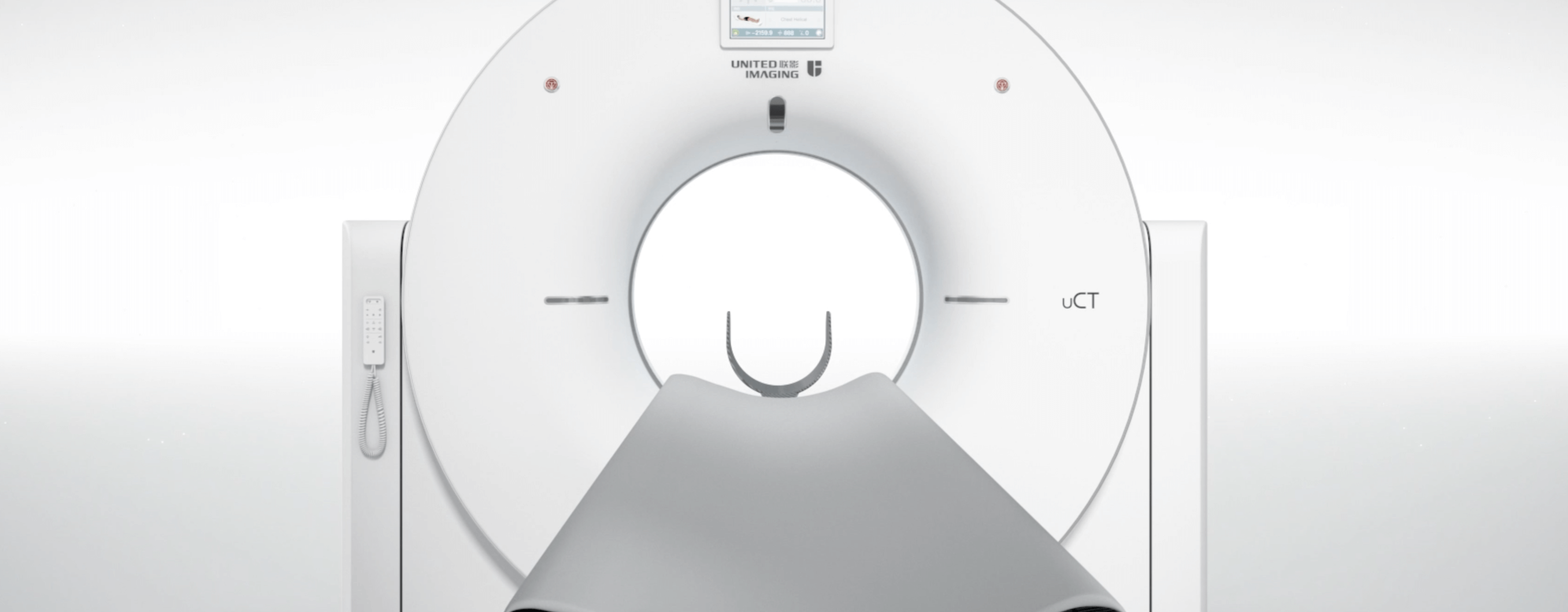
The CT scan is painless and usually takes a few minutes. The patient lies on a special table that slides into the scanner bore. It is important that the patient remain still during the examination so that the images are as clear as possible. In some cases, it may be necessary to administer contrast to help visualise certain anatomical structures.
If contrast has been administered, the patient may be asked to drink plenty of water after the examination to speed up its excretion from the body. After the examination, the patient can immediately return to normal activities, unless otherwise instructed by the physician.
How to prepare for a CT scan of the elbow?
A CT scan itself does not require prior preparation by the patient. It is important that the patient discuss any concerns and potential contraindications before the examination. On the other hand, if the attending physician has ordered a contrast-enhanced examination, appropriate preparation will be necessary.
If contrast agent administration is planned, a blood test may be required beforehand to assess kidney function (measure creatinine levels). These results will help assess whether it is safe to administer contrast. Depending on the physician’s recommendations, the patient may also be asked not to eat or drink for several hours before the examination, especially if contrast will be administered. It is usually recommended to abstain from eating for 4–6 hours before the examination, but drinking water is usually allowed. If the patient is taking medications, he or she should discuss with the physician whether these should be taken as scheduled or whether they should be discontinued before the examination.
When is it necessary to administer a contrast agent?
Not every CT scan requires contrast to be administered. Contrast-enhanced scans are often used to better visualise tumours, which may have a density similar to surrounding tissue on non-contrast CT images. A contrast-enhanced scan helps determine tumour size, shape, borders and vascularisation, which is crucial for diagnosis and treatment planning.
Contrast administration enables blood flow through tissues to be assessed, which is important in diagnosing ischaemia or assessing tumour vascularisation. If osteoarthritis is suspected, contrast can help assess the severity of the disease and the spread of the inflammation.
*ATTENTION! The information contained in this article is for informational purposes and is not a substitute for professional medical advice. Each case should be evaluated individually by a doctor. Consult with him or her before making any health decisions.


