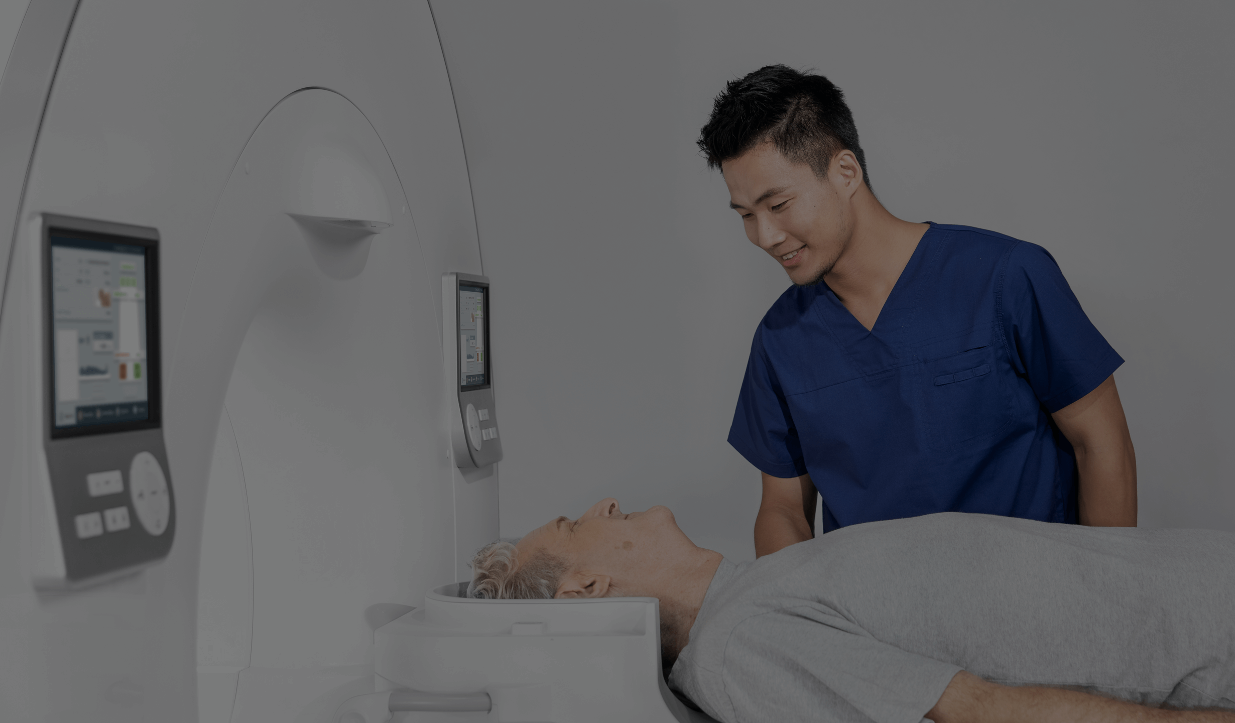
CT scan of the temporal bone pyramids
CT scan of the temporal bone pyramids is an advanced imaging study that plays a key role in the diagnosis of various diseases of the ear, nerves and bony structures of this sensitive part of the skull. Thanks to the high resolution and precision of the images obtained during CT scanning, it is possible to accurately visualise fine anatomical elements, such as the middle and inner ear structures, the auditory nerve canal, the paranasal sinuses and the complex skeletal system. This examination is particularly useful in the diagnosis of inflammatory conditions, congenital defects, cancerous lesions and injuries, enabling the physician to make an accurate diagnosis and provide appropriate treatment. Due to the exposure of the patient to X-rays, CT scans are only performed under a doctor’s referral.
What is a temporal bone pyramid?
The temporal bones (temples) are a pair of irregular bony structures that attach to the occipital bone to form the upper lateral sides of the skull. The rocky part of the temporal bone is called the temporal bone pyramid. They are an integral part of the skull and play a key role in both protecting the brain and maintaining the sensory and mechanical functions of the face and head.
The temporal bones protect the brain from mechanical trauma. Their strong structure provides solid protection for the temporal lobes of the brain, which are responsible for processing auditory and memory information. The temporal bones also act as a support for the brain's structures, keeping them in the correct position within the skull.
Another important role of the temporal bones is to cushion the movements of the lower jaw during chewing. Because of their flexibility and connections to other cranial bones, the temporal bones absorb the forces generated during biting and chewing, thus protecting the temporomandibular joints from excessive stress and damage.
The temporal bones also provide excellent support for the throat and larynx and other organs in the neck. Their position and structure help maintain the airway and support the vocal function of the larynx, allowing us to speak and breathe efficiently.
Indications for temporal bone CT scans
Because of the complicated structure of the temporal bones, a simple X-ray examination may not be sufficient. In such cases, a CT scan of the temporal bone is useful, as it allows a series of images to be taken from different angles and in different planes. This allows the doctor to get a clear picture of the patient's condition without the need for invasive procedures. Indications for a CT scan of the temporal bone pyramids include:
- evaluation of middle and inner ear structures;
- diagnosis of congenital defects;
- analysis of the extent and consequences of injury;
- suspected inflammation;
- detection of benign as well as malignant growths.
Contraindications to CT scanning of the temporal bone
A CT scan is a painless and non-invasive examination. However, there are some specific contraindications to this imaging test, such as:
- pregnancy;
- hyperthyroidism;
- renal dysfunction;
- acute reactions to the administration of contrast media.
How long does a temporal bone CT scan take?
A temporal bone CT scan usually takes 10 to 20 minutes. The scan itself takes only a few minutes, but additional time may be needed for patient preparation, such as positioning and completing documentation.
How to prepare for a temporal bone scan?
A CT scan of the temporal bone pyramids is usually performed without the use of contrast media, which means that the patient does not need to prepare for the scan. However, it is recommended that the patient arrives at the facility a short time before the appointment to complete any necessary forms or other documentation required prior to the scan. It is essential to bring a referral for a CT scan. The medical staff will also ask the patient to remove any metal objects such as glasses, pins or jewellery.
On the other hand, if the doctor has indicated that the CT scan will require the administration of a contrast agent, the patient must prepare accordingly. Contrast-enhanced CT scans require the patient to fast for at least 5 hours before the scan and to have recent creatinine and TSH tests (not older than 14 days). Patients taking metformin-containing medications who do not have significant kidney damage may continue to take these medications.
However, if a patient is taking metformin and has significant kidney damage, he or she can take the medication as planned until the examination, but should stop taking it for 48 hours after the examination and then have a creatinine test.
Computed tomography using advanced equipment from United Imaging Healthcare
A CT scan is one of the most commonly used imaging tests, especially when fast and accurate results are required. Thanks to the innovative technology developed by United Imaging, it is possible to use low doses of radiation during the examination while obtaining very high quality images (even three-dimensional) in a short period of time. This solution significantly minimises the risk of overexposure to X-rays. United Imaging’s systems are characterised by high performance and accuracy, and ensure safety during the examination.
*IMPORTANT! The information contained in this article is for informational purposes only and is not a substitute for professional medical advice. Each case should be evaluated individually by a doctor. Consult with your doctor before making any health decisions.
