Coronary CTA in the diagnosis of coronary artery disease and assessment of myocardial infarction risk
Coronary computed tomography angiography (CCTA) is an advanced imaging technique used to assess the condition of the coronary arteries that supply blood to the myocardium. It is a non-invasive method of accurately assessing the presence of atherosclerotic plaques, their characteristics and the degree of stenosis. CT is particularly useful in diagnosing coronary artery disease and assessing the risk of myocardial infarction.
What is coronary CTA used for?
Coronary CTA is mainly used to diagnose and monitor heart disease, especially coronary artery disease.
- CT can assess the presence of atherosclerotic plaque and the degree of narrowing of the coronary arteries, which can restrict blood flow to the heart. It is particularly useful in patients with symptoms such as chest pain when coronary artery disease is suspected.
- CT imaging, which can assess the type and stability of atherosclerotic plaques, helps to determine the risk of a cardiovascular event. Unstable plaques can rupture, which is a common cause of heart attacks.
- CT scan results help doctors make treatment decisions, including whether an invasive procedure (such as angioplasty or bypass surgery) is needed, or whether medication can be used.
- CT is used to monitor the progression of coronary artery disease and assess the effectiveness of treatment. It can be used to monitor changes in the condition of the vessels, such as plaque growth or stabilisation.
- CT can be used to calculate a calcium score, which reflects the amount of calcium in the coronary arteries. It is a marker of atherosclerosis and a tool for assessing cardiovascular risk, especially in asymptomatic individuals.
Indications for cardiac and coronary CT scans
The main indication is the diagnosis or exclusion of coronary atherosclerosis. Coronary computed tomography angiography is also used in patients who are being prepared for selected procedures, such as heart valve replacement, and in patients following coronary artery bypass grafting to assess the success of the procedure. Other indications include the diagnosis of arterial and venous disease, particularly the presence of aneurysms, vascular dissection, pulmonary embolism, atherosclerosis or venous thrombosis.
Contraindications for cardiac and coronary CT examinations
Cardiac CT should not be performed in pregnant women. Other contraindications arise from the need to administer contrast media. Because of their iodine content, contrast media should not be used in patients who are allergic to iodine. Contrast media can also affect renal function, so CT should not be used to diagnose patients with renal failure. Other contraindications to cardiac CT include:
- uncompensated hyperthyroidism;
- multiple myeloma;
- pheochromocytoma.
- sickle cell anaemia.
What does coronary CT scanning look like?
The examination uses X-rays and computed tomography to produce detailed, three-dimensional images of the heart’s blood vessels.
Course of the examination
- The patient usually lies on the CT scanner table. Before the scan, the patient may be given medication to slow down the heart rate, which helps to produce clearer images. Sedatives are sometimes used to help the patient relax and lie still.
- An iodine contrast agent is injected into the patient’s vein (usually in the arm) to “light up” the coronary vessels, making them more visible on CT images. Contrast enhancement makes it easier to distinguish the vessels and detect any narrowing or blockages.
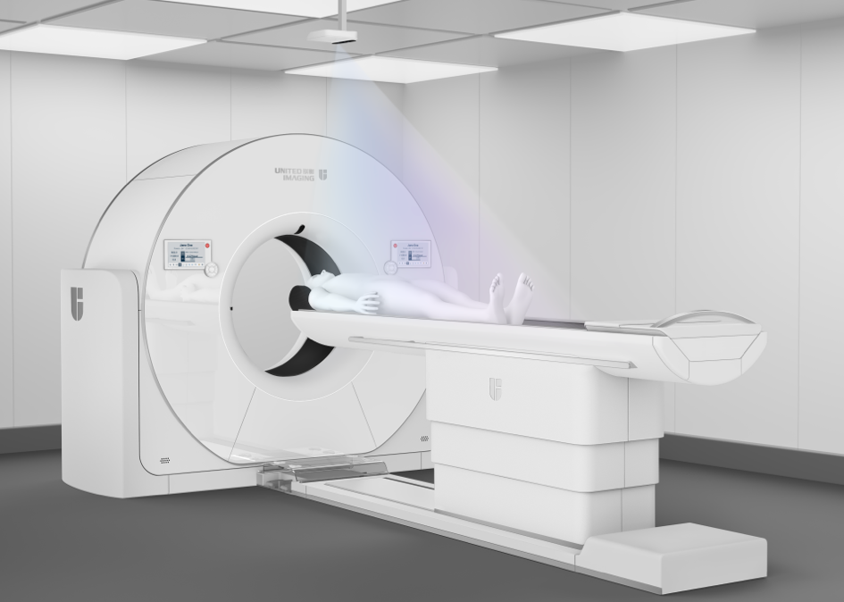
- The table slides inside the CT scanner, which rotates around the patient, taking multiple images in a short period of time. The machine scans the chest and produces precise images of the coronary arteries in different cross-sections.
- The computer combines the images obtained to create a three-dimensional image of the coronary arteries, showing any changes such as atherosclerotic plaque, narrowing or other abnormalities.
What does a coronary CT scan show?
The examination makes it possible to assess:
- the presence and extent of atherosclerotic plaques;
- the degree of narrowing of the coronary arteries;
- the type of plaque (hard, calcified, soft);
- the condition of the vessels after heart procedures such as bypass graft surgery.
How long does a coronary CT scan take?
A coronary CT scan (angio-CT of the coronary arteries) usually takes about 10–15 minutes. The scan itself is relatively quick, but the preparation, including the administration of the contrast agent and patient instruction, can extend the time spent in the scanning room to about 30-40 minutes.
How to prepare for a coronary CT scan?
Because of the need to administer a contrast agent, the patient should be fasted and should come with a referral from the doctor who has qualified the patient for the CT scan and ruled out any contraindications. The contrast agent is administered through an intravenous line (cannula). During the examination, the patient lies still on a table that moves inside the CT scanner; it is important not to move in order to avoid disturbing the images. The patient remains in voice contact with the medical staff at all times so that the examination can be interrupted if necessary. Depending on the facility, the waiting time for results is about a week.
*IMPORTANT! The information contained in this article is for informational purposes only and is not a substitute for professional medical advice. Each case should be evaluated individually by a doctor. Consult with your doctor before making any health decisions.
