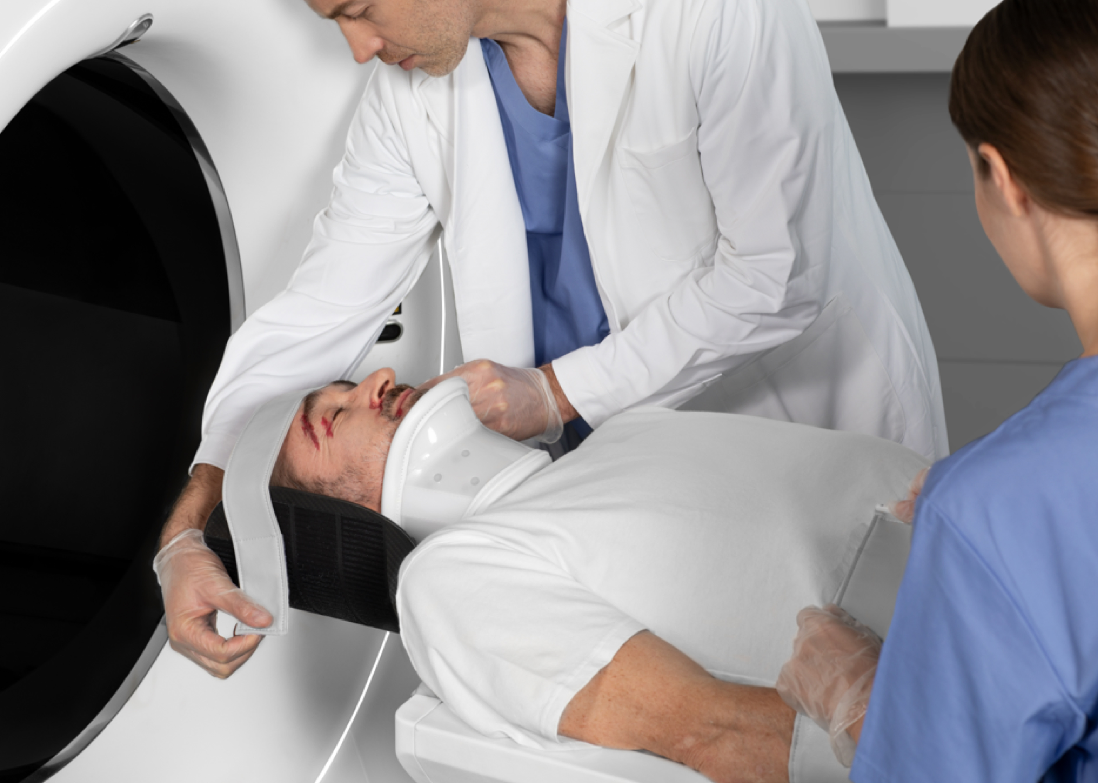
CT scan of the middle ear
A CT scan of the middle ear is an advanced imaging test based on computed tomography (CT) technology. It provides a detailed assessment of the anatomy of the middle ear and surrounding structures, such as the eardrum, bones of the ear and paranasal sinuses. The use of CT scans in otological diagnosis is an important step in understanding the complex structure of the ear, which is crucial to properly assessing its function. The high-resolution images produced allow doctors to accurately analyse various lesions.
What is the middle ear?
The middle ear is one of the three main parts of the ear, along with the outer ear and the inner ear. It is located between the eardrum and the inner ear and plays a key role in the hearing process. The middle ear is made up of the tympanic membrane (eardrum), three ossicles (bones of the ear) called the malleus, anvil and stirrup, the tympanic cavity and a canal called the Eustachian tube, which connects the tympanic cavity to the nasopharynx.
The middle ear plays a key role in the hearing process, allowing the effective transmission of sounds from the environment to the inner ear. Any disruption to its function, such as infection, injury or anatomical defects, can lead to hearing problems.
What conditions can affect the middle ear?
The middle ear can be affected by a number of conditions that can affect its function and our ability to hear. The most common problems include otitis media with effusion, where fluid accumulates in the eardrum cavity without any infection, causing a feeling of fullness in the ear and hearing problems, and otosclerosis – an inherited condition that causes abnormal growth of bone tissue around the ossicles, resulting in their immobilisation and subsequent hearing loss. In rare cases, tumours, both benign and malignant, can develop in the middle ear, causing hearing loss and other complications.
Detailed imaging of the inner structures of the middle ear allows the doctor to make a correct diagnosis and start appropriate treatment.
What is a CT scan of the middle ear used for?
A CT scan of the middle ear is used to diagnose many conditions affecting the middle and inner ear, including:
- middle and inner ear inflammation;
- aneurysms;
- tumours;
- temporal bone fractures;
- vertigo;
- hearing problems;
- degenerative diseases and other pathologies that can affect our hearing ability.
This examination is particularly useful in situations where conventional diagnostic methods, such as audiometry or otoscopy, do not provide sufficient information about the patient’s condition.
Contraindications to a CT scan of the middle ear
The main contraindications for an ear CT scan are the presence of electronic implants, such as pacemakers, cochlear implants and other electronic devices in the body that may be susceptible to interference or damage from X-rays. It should also be noted that patients with other implanted medical devices, such as neurostimulators or insulin pumps, require a thorough risk assessment prior to the scan.
How long does a middle ear CT scan take?
The duration of an ear CT depends on the imaging technique used, but the procedure is usually relatively short. The scan itself usually takes only a few minutes, but the whole appointment can take longer due to preparation and discussion of the results.
How to prepare for a middle ear CT scan?
Preparation for a CT scan of the ear is usually very simple and requires little involvement from the patient. Patients are advised to remove all metal objects from the head area, including earrings, glasses, hair clips and dentures, as the presence of metal can affect the quality of the images obtained and lead to false results.
There is no need to follow a special diet or stop taking any medication before the scan, unless the doctor advises otherwise, for example when using certain contrast media. Prior to the scan, the doctor or technician will have a brief conversation with the patient to discuss the procedure and check for any contraindications to the scan, such as implanted electronic devices or allergies to contrast media (if these are to be used).
Innovative CT scanners from United Imaging Healthcare
State-of-the-art CT scanners from United Imaging Healthcare produce the highest quality images in real time, using low doses of radiation, making the examination less stressful for the patient’s body. The advantage of CT scanning is its speed, as the required images of the inner ear structures can be obtained in a short time and after that, appropriate treatment can then be provided.
*IMPORTANT! The information contained in this article is for informational purposes only and is not a substitute for professional medical advice. Each case should be evaluated individually by a doctor. Consult with your doctor before making any health decisions.
