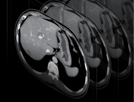
Abdominal X-ray
It is most often carried out to determine the cause of abdominal pain, nausea or vomiting. In the case of urological patients, its purpose is to visualise lesions within the urinary tract, especially calcifications of the urinary tract. A referral from a doctor is needed for an abdominal X-ray.
What is inside the abdominal cavity?
The abdominal cavity is a large space in the human body located between the chest and pelvis. It consists of the peritoneal cavity and the extraperitoneal space, which in turn is divided into three spaces. From above, it is bounded by the diaphragm, which separates it from the thoracic cavity.
Most of the abdominal cavity is filled with the organs forming the digestive system. The intestines, which occupy the largest space in the abdominal cavity, have a total length of 6 to 8 metres. The retroperitoneal space contains important parts of the urinary system, such as the kidneys and parts of the ureters.
Most common abdominal conditions
Abdominal pain is extremely common in medical practice. By analysing the nature of the pain and its location, doctors can draw conclusions about the specific ailment or organ that is the source of these symptoms.
Sudden and potentially life-threatening abdominal illnesses are referred to as “acute abdomen” in medicine. An acute abdomen is a set of symptoms associated with a variety of serious abdominal conditions that have a common characteristic in that they require immediate medical intervention.
Some of the causes of acute abdomen include:
- appendicitis;
- massive gastrointestinal bleeding;
- acute cholecystitis;
- intestinal obstruction;
- or peritonitis.
Abdominal pain is often of the referred (or reflective) kind, meaning that pain in a particular area may be caused by a pathological condition in another area. This is due to the common innervation of many organs and structures. A gastroenterologist – a doctor who treats abdominal conditions – may refer the patient to another specialist depending on the diagnosis.
Indications for an abdominal X-ray
Abdominal X-ray is performed urgently when obstruction or perforation of the gastrointestinal tract is suspected. Especially if there are symptoms such as fever, rapid heartbeat, vomiting blood or very severe abdominal pain, an X-ray examination is usually performed in a hospital emergency department (ED).
The examination is also carried out for diagnostic purposes to confirm or rule out a particular condition, e.g. in the diagnosis of diseases of the digestive or urinary tract.
Abdominal X-ray – contraindications
An abdominal X-ray is a painless and safe examination, but it is not recommended in certain situations. An abdominal X-ray is not indicated for:
- pregnant women;
- young patients;
- women of childbearing age in the first 12 days of the menstrual cycle (unless the doctor decides otherwise).
During the examination, X-rays are used, which, although the dose absorbed during a single examination is not very high, are not completely harmless to the human body. Therefore, although a single exposure does not result in a high radiation dose, frequent X-ray examinations should be avoided.
What does the examination look like?
During an abdominal X-ray, an electroradiologist takes a series of X-rays using special equipment. The key is to strictly follow staff instructions and avoid movement during the exposure. If requested by the technician, the patient should also hold his or her breath for a few seconds to avoid blurring the image. The results of the examination are discussed during the consultation with the doctor.
An abdominal X-ray usually takes a few minutes. The examination may be prolonged if additional images are required to get a more accurate picture.
How to prepare for an abdominal X-ray?
X-ray evaluation may be more difficult if large amounts of gas are present in the intestines, so proper preparation for the examination is required.
The patient should bring the following documents for the abdominal X-ray examination:
- a current referral from a physician;
- results of previous diagnostic tests (impressions and CDs);
- discharge summary from the hospital, if the examination was recommended in the discharge summary;
- ID card or the child’s medical records in case of minors.
The day before the examination, a semi-liquid diet is recommended, and the patient should avoid fruits, vegetables, sweets and beverages such as fruit juices and sodas. The patient should also take an anti-foaming agent, such as simeticone, on this day.
*IMPORTANT! The information contained in this article is for informational purposes only and is not a substitute for professional medical advice. Each case should be evaluated individually by a doctor. Consult with your doctor before making any health decisions.


