X-ray of the spine
X-ray of the spine is a key examination that enables the bones that make up the spine to be visualised. This examination makes it possible to detect posture problems, injuries, degenerative changes and inflammation. It is one of the diagnostic tests used when cancer and spinal metastases are suspected. It is also performed to determine the cause of pain in a specific part of the spine.
Depending on the ailment, the following X-ray examinations of the spine can be performed:
- X-ray of the lumbosacral spine;
- X-ray of the cervical spine;
- X-ray of the thoracic spine.
A doctor’s referral is required for a spinal X-ray examination, irrespective of whether it will be funded by the National Health Service or paid privately.
Diseases of the spine
Some of the most common spinal conditions include:
- degenerative spinal disease;
- discopathy;
- ankylosing spondylitis;
- osteoporosis;
- spinal stenosis.
Causes of spinal pain syndromes are mainly identified on the basis of a detailed examination by a physician or physiotherapist. If there are alarming symptoms, this examination may be supplemented by additional tests, such as a spinal X-ray.
How are spinal diseases treated?
Treatment is usually conservative. In cases without alarming symptoms, in addition to painkillers and anti-inflammatory drugs, the main recommendation is to introduce changes to daily routine. It is important to improve workplace ergonomics, make short exercise breaks during sedentary work, engage in daily physical activity (such as walking, swimming or cycling), wear comfortable shoes, and sleep on a mattress that fits the patient’s body weight.
Indications for X-ray examination of the spine
X-ray examinations of the spine are performed only for important reasons. The main indications for this examination include:
- degenerative changes of the spine;
- spinal injuries;
- posture problems;
- suspected developmental defects;
- suspected cancer metastases to the spine;
- spondyloarthropathies (arthritis involving the joints of the spine);
- headaches and dizziness.
X-ray examinations of the spine are performed for a variety of reasons and provide one of the diagnostic methods of assessing posture problems and their severity. X-rays also make it possible to identify inflammatory and degenerative changes of the spine.
This examination is also recommended for patients who have suffered spinal injuries, as it can assess whether any fractures, dislocations or vertebral displacements have taken place.
Contraindications to the examination
The only contraindication to a spinal X-ray examination is pregnancy. During the examination, the patient is exposed to X-rays, which are particularly harmful to the foetus. For this reason, in pregnant women, X-ray and other examinations that involve radiation are only performed in life-threatening situations, when they are necessary for diagnosis. In other cases, imaging studies such as ultrasound or MRI are recommended.
Many people question whether X-ray examinations are harmful to health. X-rays are not completely neutral to the human body, so this type of examination should only be performed where required by the health situation.
Caution is also required when performing spinal X-rays in children – although young age is not an absolute contraindication, these examinations should not be performed too frequently in young patients.
How to prepare for a spinal X-ray?
X-ray examination of the spine usually does not require special preparation from the patient, with the exception of lumbosacral spine X-ray. In this case, the patient’s intestines need to be cleansed of faecal masses and gases that may interfere with image readability. For the lumbosacral X-ray, the patient should be fasting. Where possible, this examination should be performed in the morning.
On the day of the appointment, it is advisable to wear comfortable, two-piece clothing to make it easier to undress for the examination. Patients (especially female patients) feel more at ease if they only need to undress from the waist up.
What does an X-ray examination look like?
The duration of the examination depends on the number of images to be captured and usually ranges from 5 to 15 minutes from the time the patient enters the laboratory.
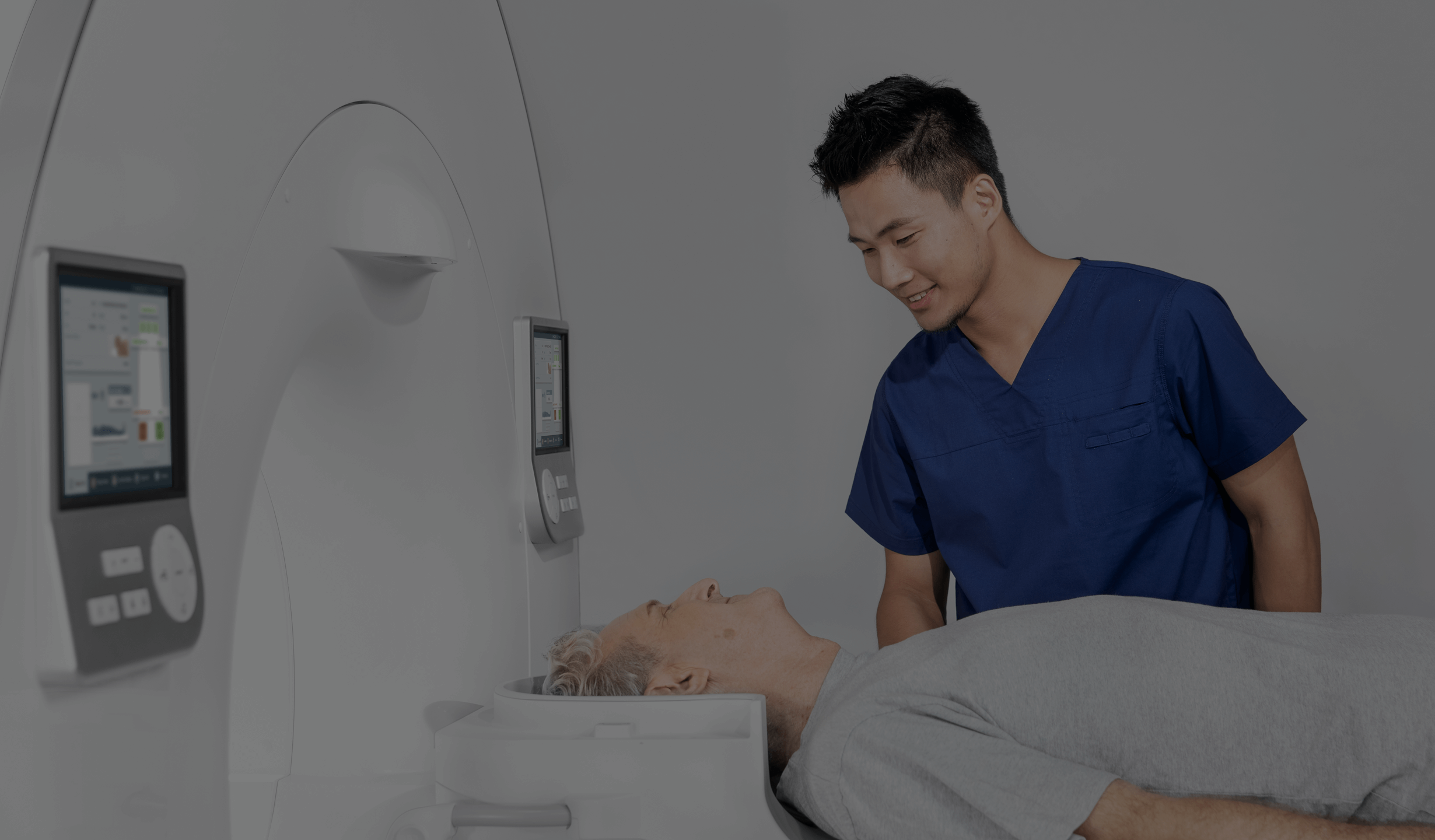
It is performed in a specialised laboratory where the patient must follow the electroradiologist’s instructions. In order to perform the examination, the section of the spine to be examined must be exposed, and clothing and all jewellery must be removed from the area to be examined. The technician will indicate the proper positions to the patient – possible image projections include e.g. the lateral, anterior-posterior, oblique or functional projection (in flexion and extension). After the examination has been completed, the patient can immediately return to daily activities.
*IMPORTANT! The information contained in this article is for informational purposes only and is not a substitute for professional medical advice. Each case should be evaluated individually by a doctor. Consult with your doctor before making any health decisions.


