Adenoid X-ray
Adenoid X-ray is a specialist radiological examination that is often performed in children to assess the size and degree of adenoid (pharyngeal tonsil) hypertrophy. This is particularly important in diagnosing problems related to, for instance, nasal breathing, recurrent respiratory infections and snoring.
What is the adenoid?
The adenoid is a lymphatic structure located in the upper part of the throat, near the nasopharynx. It is a part of the immune system that helps protect the body from infections, especially during childhood when the immune system is still in its development stage.
Unlike the palatine tonsils, which are visible when the mouth is opened, the adenoid is located higher up, behind the nasal cavity, and as a result it is not visible during a routine throat examination. It is most active in the first years of a child’s life, and usually shrinks with age, often disappearing completely by adolescence.
Adenoid conditions in children
The most common conditions affecting the adenoid in children include the following:
Adenoid hypertrophy
Adenoid hypertrophy mainly affects children between the ages of 3 and 7, although it may also occur in older children. If the examination confirms adenoid hypertrophy, the doctor may recommend various treatment options ranging from observation and conservative treatment to surgery (adenotomy) to remove the hypertrophied adenoid.
Adenoid hypertrophy can lead to various health problems. Because the adenoid is located near Eustachian tube openings, its enlargement may lead to ear problems, such as recurrent middle ear infections or chronic ear and upper respiratory tract infections. Adenoid hypertrophy may also block airflow through the nose, causing breathing difficulties, snoring and even sleep apnoea.
Chronic adenoiditis
Chronic adenoiditis is a condition involving long-term inflammation of this lymphatic structure. It is a common problem in children and can lead to a number of unpleasant symptoms and complications affecting the child’s quality of life and normal development.
Chronic adenoiditis can be caused by various factors, including frequent viral or bacterial infections, allergies, air pollution or even cigarette smoke.
Recurrent middle ear inflammation
Recurrent middle ear inflammation (otitis media) is a condition involving multiple episodes of inflammation of the middle ear – the area behind the eardrum where the auditory ossicles are located. This is a common ailment in children, especially those of preschool and early school age. Frequent ear infections can lead to serious complications, including problems with hearing and speech development.
Treatment often includes antibiotic therapy, and if infection recurs frequently, it may be necessary to place a drain tube in the tympanic membrane and remove the adenoid.
How to prepare for an adenoid X-ray?
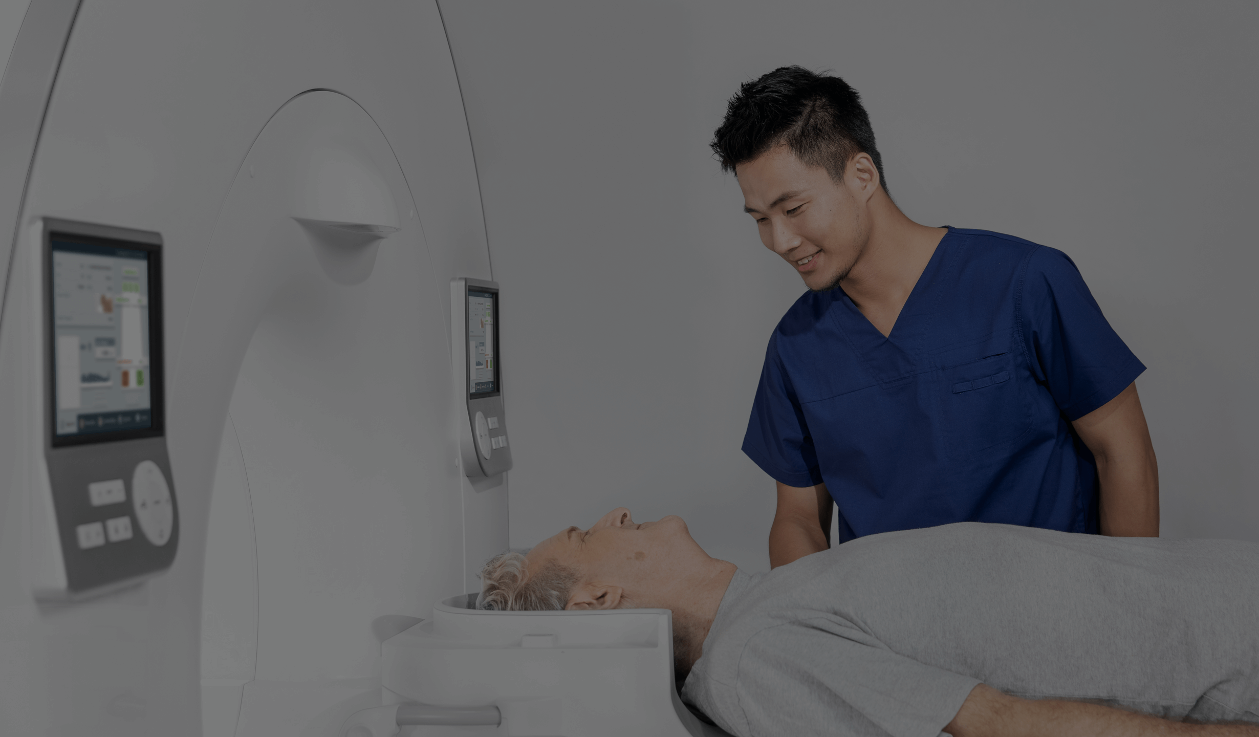
No special preparation is required. However, it is important that the child be calm and cooperative during the examination. Sometimes parents are present in the room to make the child feel safer.
Before the examination, any jewellery or metal objects should be removed from the child’s head and neck area so that they do not interfere with the X-ray image. Medical staff will explain to the child and parents exactly what will happen and what steps will be taken. It is important that the child remains still, as movement can affect the quality of the image.
It should be remembered that X-ray examinations are widely used and are safe if doses are properly controlled. Medical staff take care to minimise radiation exposure to ensure the safety of patients, including children.
How is an adenoid X-ray performed?
Adenoid X-ray is a quick and non-invasive examination. During the examination, the child is asked to position the head in a certain way to ensure the best view of the adenoid (pharyngeal tonsil). Subsequently, an X-ray is taken to assess the size of the adenoid and its possible effect on airflow through the airways.
The radiation dose used during this examination is minimal, but the radiology technician and parents (if present in the room) can still be equipped with special protective shielding to minimise radiation exposure.
Once the image is taken, the radiologist evaluates it for the presence of an overgrown adenoid. The examination makes it possible to assess whether the adenoid is causing a partial or complete blockage of the nasopharynx.
The radiologist prepares a report, which is then forwarded to the attending physician. Based on the X-ray results, the physician will decide on further treatment, including the possible need for ENT consultation or surgery.
If there are any doubts whether an adenoid X-ray is necessary, it is always advisable to consult an ENT specialist, who will assess whether this examination is necessary and whether its results may have a significant impact on further medical management.
Alternatives to X-ray
Where there are doubts as to the diagnosis or a more detailed picture is needed, the physician may order the following examinations:
- Nasopharyngeal endoscopy – an examination with an endoscope that allows the adenoid (pharyngeal tonsil) to be viewed directly and its size and condition to be assessed.
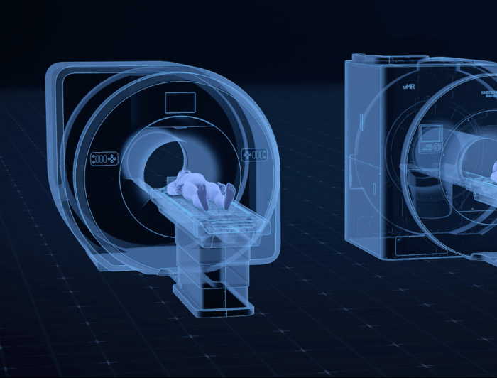
- Magnetic resonance imaging (MRI) or computed tomography (CT) – these are more advanced imaging modalities that can be used in special cases, but are not usually needed to evaluate the adenoid.
*ATTENTION! The information contained in this article is for informational purposes and is not a substitute for professional medical advice. Each case should be evaluated individually by a doctor. Consult with him or her before making any health decisions.


