What is radiovisiography?
Radiovisiography (digital dental radiography) is an advanced medical imaging technique that uses digital technology to produce X-ray images. Unlike traditional X-rays, which require the use of film, radiovisiography allows images to be produced directly in digital form. This allows the image to be quickly viewed on a computer screen, and it can also be easily stored, analysed and transmitted.
Digital radiography is widely used in dentistry to accurately diagnose problems with teeth, roots and jawbones. The technology is not only more accurate than traditional X-rays, but also involves less radiation exposure for the patient.
What is radiovisiography in dentistry?
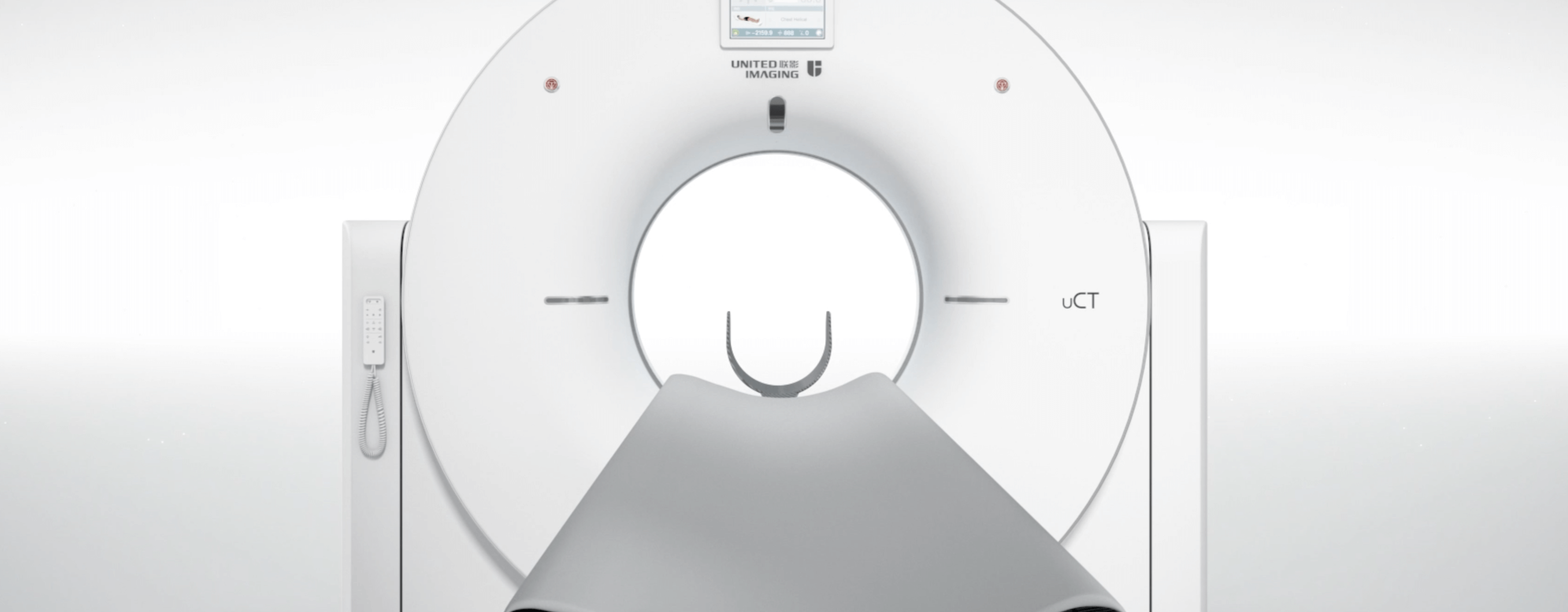
Radiovisiography (RVG) in dentistry is an advanced digital X-ray imaging technique used to diagnose and monitor the condition of teeth and periodontium. Using RVG, dentists can obtain high quality images of a tooth in seconds, enabling them to more accurately assess roots, bone, caries and the effectiveness of endodontic or implant treatment. United Imaging Healthcare provides the highest quality medical equipment to support leading medical facilities.
Advantages of RVG in dentistry
- Lower radiation doses – RVG requires a much lower radiation dose than conventional X-rays, protecting the health of the patient.
- Speed – the image appears almost instantly on the computer screen, reducing appointment time and allowing immediate analysis.
- Accuracy – the digital image can be magnified and its contrast and brightness can be adjusted, helping the dentist to see fine details.
- Easy storage and archiving – digital images can be stored on the computer, making it easy to monitor treatment progress and share images with other specialists.
How to prepare for an RVG?
Preparing for an RVG is straightforward and does not involve many steps, but there are a few recommendations that can help to ensure a smooth and uninterrupted examination. Firstly, all metal objects such as earrings, necklaces, glasses, hairpins and removable dentures should be removed from the head and neck area before the examination. Metals can interfere with the examination and cause image artefacts that can make the diagnosis difficult.
The patient should maintain good oral hygiene before the examination. Removing all food particles and chewing gum will help the dentist get a clear picture and make it easier to interpret the results. The patient should remain still during the exposure. Even minimal movement can blur the image. The correct head position will be determined by the dentist or dental assistant.
The patient wears a lead apron to protect the rest of the body from radiation. It is important that the apron fits snugly, especially around the neck and torso. Pregnant women should inform their doctor of their condition. Although the dose of radiation used in RVG is very low, exposure to X-rays during pregnancy should be kept to a minimum. Radiovisiography does not require any dietary restrictions or taking any medication before the test.
What conditions can be diagnosed using radiovisiography?
RVG produces high quality digital images that allow doctors to detect even small lesions, contributing to early diagnosis and effective treatment.
- Caries – RVG can detect caries, including early lesions that may not be visible to the naked eye, especially in hard-to-reach areas such as interdental spaces.
- Periapical lesions – the examination helps to diagnose inflammatory conditions present around the roots of teeth, such as abscesses or cysts. These are visible as lesions around the root tip and indicate inflammation or infection.
- Pulpitis and pulp disease – RVG allows the condition of the tooth's pulp to be assessed and helps to identify lesions caused by infection or inflammation, which is important when planning root canal treatment.
- Periodontal lesions – radiovisiography can assess the condition of the bone and periodontium (the structures that support the teeth), which is crucial in diagnosing periodontal diseases such as periodontitis.
- Injuries and fractures – RVG can help detect tooth root fractures or other injuries, such as enamel and periapical bone fractures that may require specialist treatment.
What does a radiovisiography examination look like and how long does it take?
Digital radiography is a fast, painless and minimally invasive diagnostic procedure in dentistry that takes only a few minutes to complete.
What does a radiovisiography examination look like?
The patient sits in the dental chair and the dentist or dental assistant prepares the patient for the examination; a protective apron with a lead insert is put on the patient to minimise exposure of other parts of the body to radiation. A digital sensor - a small device that looks like a plastic plate - is then placed in the patient's mouth. The sensor is placed in the appropriate position depending on which teeth or structures need to be scanned; it captures the radiation and converts it into a digital image.
The dentist positions the X-ray unit close to the patient's face and points it at the sensor. The patient must remain still, as even the slightest movement can affect the quality of the image. Radiation is emitted for a very short time (a fraction of a second); during the exposure, the sensor generates the X-ray image and immediately transmits it to the computer.
The image is immediately displayed on a computer screen, where the dentist can enlarge it, adjust its contrast and perform a detailed analysis. The image can be stored in the system for comparison with future images, allowing the patient’s health to be monitored over a longer period of time.
Duration of the examination
A single RVG image takes approximately 5-10 minutes in total, including patient preparation, sensor placement, exposure and preliminary image analysis. The radiation exposure itself is very short – literally a fraction of a second. Radiovisiography is fast, convenient and effective, which is why it is widely used in everyday dental practice.
*IMPORTANT! The information contained in this article is for informational purposes only and is not a substitute for professional medical advice. Each case should be evaluated individually by a doctor. Consult with your doctor before making any health decisions.
