MR arthrography
Arthrography is an imaging test performed using magnetic resonance imaging. It involves injecting contrast into the examined joint and performing an MRI scan. The test can be performed on any joint to which contrast can be administered, such as the knee, hip, shoulder, elbow and ankle joints.
Arthrography is the third type of joint examination alongside ultrasound or X-ray. It allows non-invasive examination of the source of pain, type of damage or degree of degeneration. The additional administration of contrast allows doctors to see damage or conditions in greater detail.
Indications
The main indications for arthrography are damage and injuries to articular cartilage, menisci, articular capsules, tendons and ligaments. Therefore, contrast-enhanced examination is performed primarily for the shoulder, hip and knee joints, which are the most commonly affected by damage. Less commonly, arthrography is also used to examine the wrist, ankle and elbow joints.
Due to the different loads to which they are subject, different joints require diagnostics due to different indications.
Arthrography of the shoulder joint
- painful shoulder syndrome
- unstable shoulder joint
- complex ligament damage
- rotator cuff injuries
Arthrography of the hip joint
- damage to the acetabular labrum
- damage to the articular capsule
Arthrography of the elbow joint
- joint ligament damage diagnosis
What does MR arthrography look like?
Arthrography is a diagnostic procedure that involves the administration of a contrast agent prior to an MRI scan. Contrast is administered either intravenously or directly into the joint.
The examination is performed in the following planes:
- transversal
- coronal
- sagittal
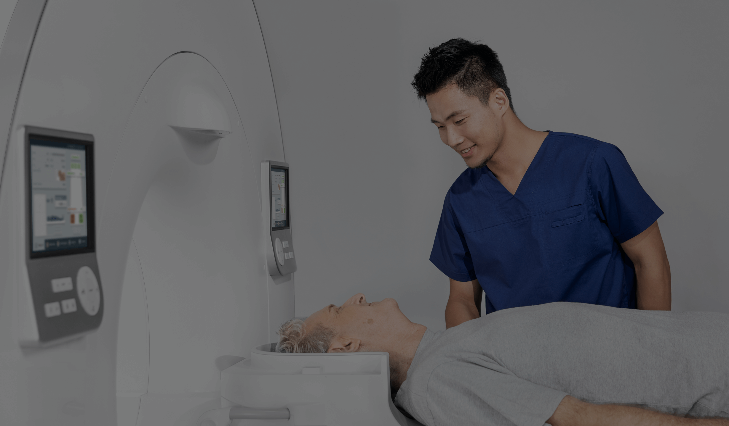
The patient lies down in the scanner bore and must remain motionless for the entire scan. Depending on the joint being examined, the entire procedure can take from 30 minutes to as much as an hour.
The examination itself is painless, but to improve comfort, an anaesthetic is first administered into the joint cavity. As a result, an arthrographic examination is not really painful and most patients tolerate it well.
More discomfort may result from a feeling of fullness of the joint capsule, which gradually subsides within 24 hours after the examination. The contrast agent is excreted from the body with urine within 48 hours. The puncture site through which the contrast agent is administered requires no special care, and the dressing can be removed after a few hours. Temporary tenderness may occur at the injection site.
How to prepare for MR arthrography?
Proper preparation for a contrast-enhanced MRI scan is crucial for both patient safety during the examination and for patient comfort.
The patient should fast for at least 6 hours before the examination. If the patient regularly takes certain medications, he or she should consult the doctor about taking them before the examination.
Up-to-date blood tests are required as well – in particular, creatinine level, full blood count and electrolyte levels (concentrations of sodium, potassium, calcium, chlorides, iron, magnesium phosphates in the blood).
If the patient has metal implants, which may be a contraindication to the scan, medical staff should be informed in advance. It is recommended that the patient arrive at least 30 minutes before the examination.
Arthrography – contraindications
As with any examination, there are several contraindications to arthrography, although not all of them are absolute. It is important to remember that the final decision whether to carry out the examination is always made by the doctor, taking into account the patient’s individual situation and assessment of his or her health.
The main contraindications include allergy to the contrast agent, pregnancy in the case of women, cancer, heart failure, stomach ulcers, chronic kidney failure, hypertension or metal implants in the body, such as joint replacements, pacemakers, dental implants or bone stabilising implants.
*IMPORTANT! The information contained in this article is for informational purposes only and is not a substitute for professional medical advice. Each case should be evaluated individually by a doctor. Consult with your doctor before making any health decisions.


