Magnetic resonance imaging of the adrenal glands
Magnetic resonance imaging (MRI) is one of the most versatile tools in medicine, allowing non-invasive examination of anatomical structures of organs. Adrenal MRI is a safe and accurate diagnostic method that can detect abnormalities in the adrenal glands. This examination can be used to determine with high probability whether a lesion is benign or malignant. This method of diagnosis is completely painless and extremely safe.
What are the adrenal glands?
The adrenal glands are organs located above the kidneys, adjacent to them, which are present on both sides of the body, just like the kidneys. They consist of two parts: the inner part, called the medulla, and the outer part, called the adrenal cortex. The adrenal cortex produces three groups of hormones: glucocorticoids (GCs), mineralocorticoids (MCs) and androgens.
Adrenal gland disorders
Adrenal gland disorders include:
- adrenal insufficiency, a condition in which the adrenal glands produce inadequate amounts of hormones (Addison’s disease);
- Cushing syndrome (a disease associated with excess glucocorticoids);
- Conn’s syndrome, secondary hyperaldosteronism, hypoaldosteronism (diseases associated with mineralocorticoid secretion disruptions);
- disorders associated with androgen excess (e.g., hirsutism, virilisation, androgen-secreting adrenal tumours);
- accidentally detected adrenal tumours (so-called incidentalomas);
- adrenal cancer;
- pheochromocytoma.
MRI can also detect adrenal gland haematomas, which may be the result of trauma or other conditions. However, it is important to note that this examination cannot directly diagnose endocrine disorders. Nevertheless, it helps doctors identify lesions in the adrenal glands that may affect the production of hormones (aldosterone, cortisol or adrenaline).
Magnetic resonance imaging of the adrenal glands – indications
Primary indications for adrenal MRI include:
- Suspected adrenal tumours, especially when there are symptoms that suggest this condition, such as hypertension, abdominal pain, hormonal changes (such as Cushing syndrome or pheochromocytoma symptoms). This examination is often recommended to accurately assess the size, location and possible nature of the lesions. It should be remembered that adrenal tumours often do not cause obvious symptoms and may be detected incidentally.
- Monitoring adrenal tumours in patients with previously diagnosed lesions or suspected cancer.
- In cases of difficulty controlling hypertension or suspected secondary causes of hypertension, such as hormone-secreting tumours (e.g., aldosteroma, pheochromocytoma), adrenal MRI may be recommended to assess the condition of adrenal glands.
- Suspected adrenal hyperplasia (overgrowth) or cysts.
- Preparation for surgical removal of adrenal tumours.
Contraindications to magnetic resonance imaging of the adrenal glands
Contraindications to an MRI examination include:
- metal orthopaedic implants;
- a pacemaker;
- neurostimulators (e.g., analgesic or brain neurostimulator);
- metal brain aneurysm clips;
- some artificial heart valves;
- pregnancy (first trimester); in the second and third trimesters, pregnancy is a relative contraindication;
- extensive tattooing in the examined area;
- fear of enclosed spaces (claustrophobia);
The presence of metal fragments such as shot, bullet fragments or iron filings is also a problem. This is because the movement and heating of metal fragments may damage surrounding tissues, and interference with the operation of electronics could pose an even greater threat to the patient.
Other possible contraindications to adrenal MRI include:
- a recent biopsy – haematomas and healing processes may cause the interpretation of results to be erroneous;
- difficulty keeping still for long periods of time – for example, due to Parkinson’s disease or nervous tics;
- excessive patient weight – the MRI scanner has a limited load capacity.
How to prepare for the examination?
All metal objects should be removed, and jewellery and hair ornaments should be taken off. The patient should appear preferably 30 minutes before the test, fasted. If the examination is to take place in the evening, the patient should eat easily digestible foods throughout the day.
After consulting the doctor, regular medications should be taken at usual times with a little water.
A rectal glycerin suppository may be administered the day before to cleanse the digestive tract of food residues and gas.
How long does an adrenal MRI take and what does it look like?
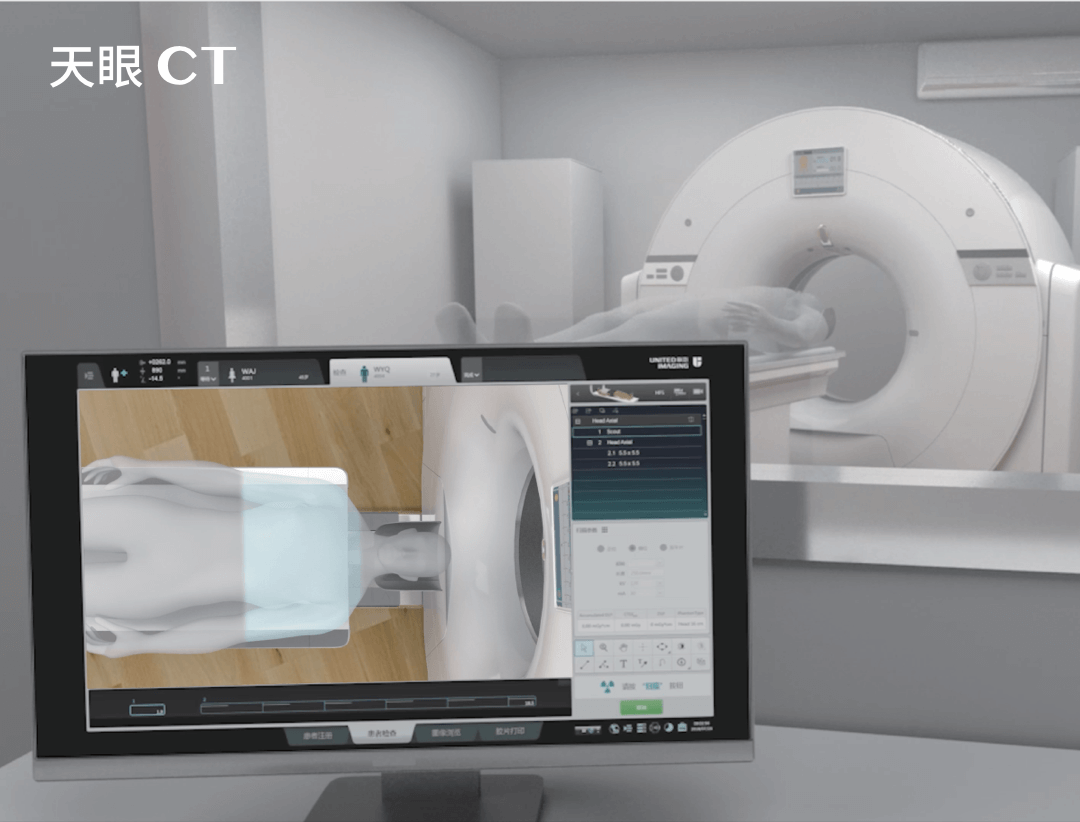
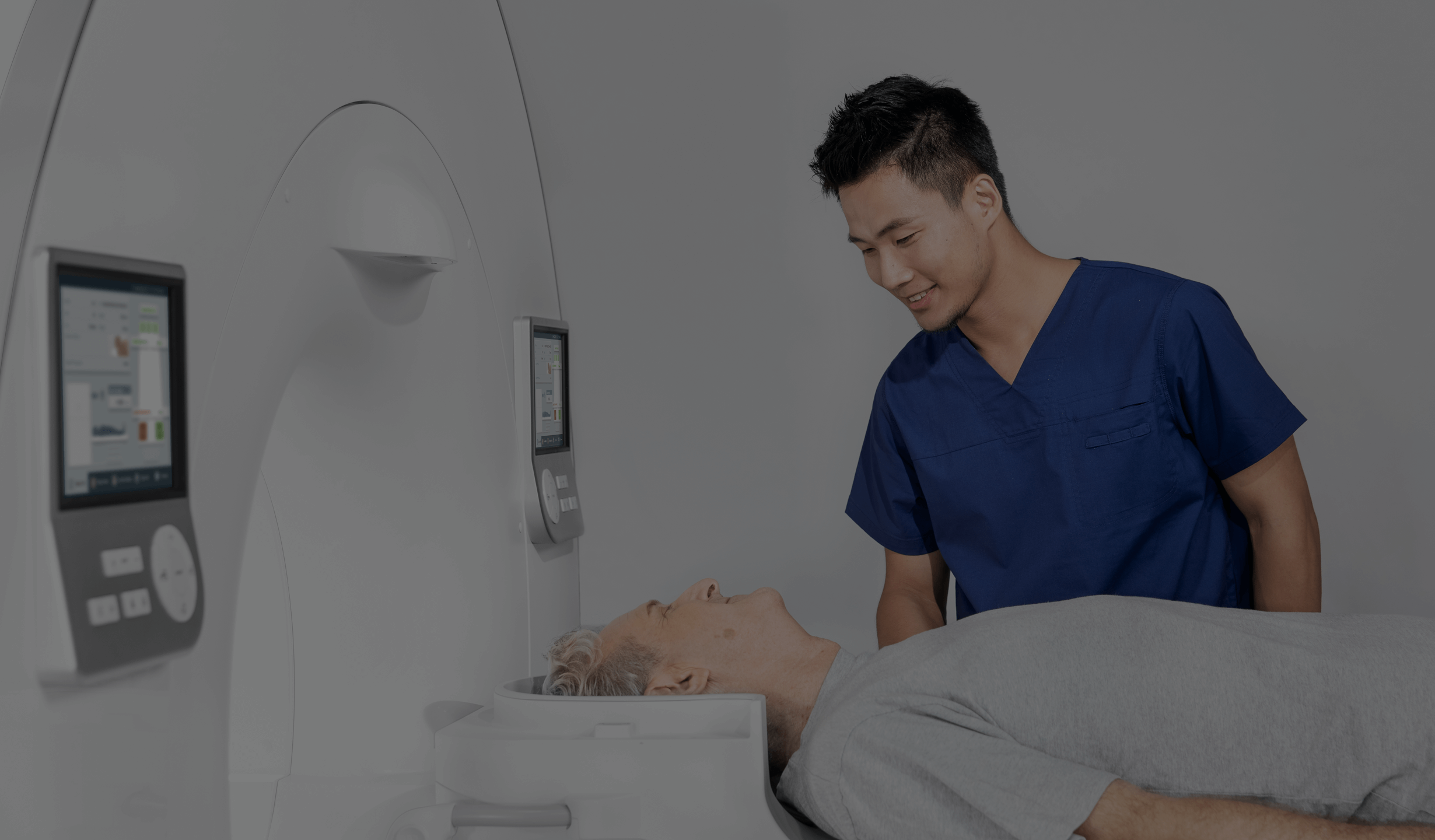
MRI examination of the adrenal glands can be performed while the patient is clothed. It is important that the patient’s clothing does not contain any metal parts. The patient lies down on a special table that automatically slides inside the scanner. The patient should remain motionless during the entire scan, as any movement can blur the image.
The person supervising the examination is in a separate room, but is able to communicate with the patient via intercom.
After the examination, the patient can immediately return to daily activities, such as eating or driving.
However, if the examination was performed using contrast or the patient is claustrophobic and sedation was required, the patient must remain under medical observation for 30 minutes after the procedure. After contrast administration, an allergic reaction can occur, and thus the patient should wait some time in the facility so that he or she can be helped quickly in an emergency.
After the administration of sedatives, a person’s concentration and reflexes will be impaired for the next few hours. The patient should not drive any motor vehicles during this time.
The duration of an adrenal MRI scan may vary depending on the specifics of the procedure, but usually ranges from 30 to 60 minutes. The patient must remain still during the examination to ensure the highest quality images. Medical staff often communicate with the patient via intercom, letting him or her know when he or she can breathe or move.
*IMPORTANT! The information contained in this article is for informational purposes only and is not a substitute for professional medical advice. Each case should be evaluated individually by a doctor. Consult with your doctor before making any health decisions.


