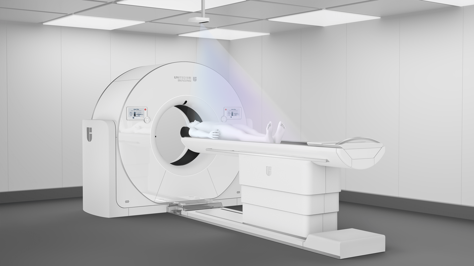
Guide to MRI for claustrophobic patients
As the use of MRI in diagnostics increases, more and more people are undergoing the procedure, including patients who suffer from claustrophobia. Claustrophobia is the fear of enclosed spaces. This fear is particularly intense in small, low and confined spaces, such as the inside of an MRI scanner. Does this phobia prevent a patient from having an MRI scan, or do medical staff have special methods to make the scan more comfortable?
What is claustrophobia and how does it manifest itself?
Claustrophobia is the fear of being in small, enclosed spaces. It is a type of specific phobia or irrational fear that causes severe anxiety and distress. Claustrophobia can be experienced in both small and large spaces that are perceived as “closed” or “confined” - such as lifts, small windowless rooms, tunnels, aeroplanes and even crowded rooms.
Claustrophobia can cause both physical and psychological symptoms. he most common physical symptoms can include a faster heartbeat, difficulty breathing, excessive sweating, shaking of the hands or body, dizziness or a feeling of weakness, nausea or a feeling of wanting to run away. Psychological symptoms may include feelings of panic and intense fear, a strong desire to leave the enclosed space, fear of losing control or “drowning” in the situation, or fear of suffocation or being trapped.
The factors that cause claustrophobia are not fully understood, but may include traumatic experiences, genetics and environmental conditions. Treatment for claustrophobia often involves cognitive behavioural therapy (CBT), relaxation techniques and, in some cases, exposure therapy, which allows the patient to gradually get used to situations that cause fear.
Is claustrophobia an absolute contraindication for an MRI scan?
In addition to contraindications such as certain cochlear implants, stents, vascular clips, defibrillators, pacemakers or neurostimulators, claustrophobia is also listed as a contraindication, particularly if the patient suffers a significant attack during the scan. During the examination, the patient must lie motionless in the MRI bore and his or her body gradually slides into the scanner. Without proper preparation, such an environment can trigger an attack. Uncontrolled shaking or even hyperventilation can affect the resulting images.
What does an MRI scan look like for people with claustrophobia?
The situation is not hopeless. Given the significant percentage of people who suffer from claustrophobia, medical professionals have developed several proven methods to make the scan as comfortable as possible for the patient.
Claustrophobia-friendly equipment
One possible solution is to enlarge the bore of the scanner in which the patient is placed. Even an extra 10 cm in diameter can make a difference. Although this may not seem like a big change, the extra space makes patients feel more comfortable and less likely to interrupt the examination. To meet these needs, United Imaging Healthcare offers a range of scanners with customisable gantry diameters.
Properly trained medical staff
An important part of the solution is the proper training of the medical staff and their attitude towards the patient. Showing interest, empathy and concern – even if acquired through training – can create an atmosphere of safety that can help to reduce symptoms of claustrophobia to the point where the examination can proceed without interruption. It is equally important that the patient is properly informed about what to expect and how to try to reduce their anxiety.
Preparing a claustrophobic patient for the examination
Patients who are claustrophobic can benefit from instructions on how to control their reactions. It is also a good idea for the patient to learn and practise some anxiety management techniques before an examination such as an MRI or CT scan. These include concentrating on regular, deep breathing; distracting yourself from the stressful situation by focusing on a pleasant memory or object; closing your eyes to avoid seeing the confined space around you; and repeating to yourself that the anticipated threat is not real.
Performing the examination under sedation
If relaxation techniques and tranquillisers do not have the desired effect, the examination can be carried out under anaesthesia or sedation. Pharmacological agents allow the patient to achieve a deep state of relaxation and even fall asleep during the examination. In such cases, an anaesthetist is present to decide beforehand on the choice of substance, its dose and method of administration (oral, inhalation or intravenous). The decision will depend on the patient's general state of health and any contraindications, such as allergies or interactions with medications the patient regularly takes.
*IMPORTANT! The information contained in this article is for informational purposes only and is not a substitute for professional medical advice. Each case should be evaluated individually by a doctor. Consult with your doctor before making any health decisions.
