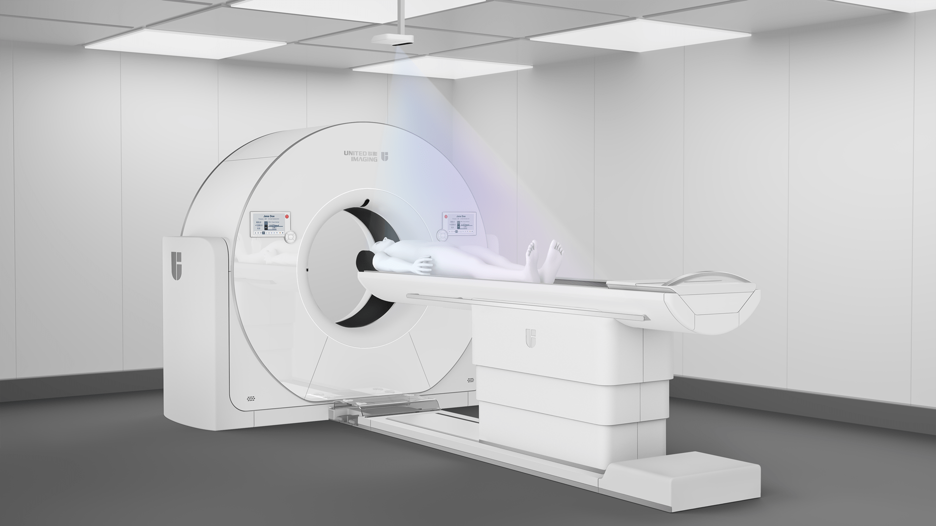
Magnetic resonance imaging of the orbits
Orbital MRI is an advanced imaging study that is used to evaluate anatomical structures within the orbits, such as muscles, nerves, blood vessels and surrounding tissues. It is a non-invasive procedure that uses a strong magnetic field and radio waves to produce detailed images of tissues. It is often used as an adjunct to other diagnostic tests, such as ultrasound and CT scans.
What is an orbit?
The orbit (eye socket) is a cavity in the skull made up of bony tissue that surrounds and protects the eyeball. It contains optic nerves, muscles, blood vessels, tear glands and fatty tissue. This complex system forms a special environment that can be examined by MRI in certain situations.
What can an orbital MRI show?
Magnetic resonance imaging can accurately assess the location and size of foreign bodies (such as shards of glass or wood) in the eye, identify and localise tumours and tumour infiltrates, show damage to the myelin sheath of nerves, detect inflammation, diagnose certain types of glaucoma, assess increased intracranial pressure, and identify blood clots in the retinal blood vessels.
Orbital MRI – indications
Orbital MRI may be recommended in a variety of situations, particularly when the cause of recurrent or chronic eye pain needs to be determined, such as inflammation, tumours or other conditions within the orbit; evaluating the optic nerves, myelin and other structures of the eye to detect possible pathology when a patient is experiencing loss or disturbance of vision or deterioration of visual acuity; diagnosing orbital tumours, both benign and malignant, and assessing their size, location and characteristics; identifying and evaluating cystic lesions that may cause swelling, pressure on ocular structures or pain, as well as trauma and inflammation.
Magnetic resonance imaging of the orbits – contraindications
Although orbital MRI is considered to be a safe diagnostic method, in some cases it can be difficult to perform. Patients with metal implants should be cautious. This includes, but is not limited to, patients with dental fillings or braces (a certificate from the orthodontist regarding the type of materials used is required), vascular clips and valves, pacemakers, implants, insulin pumps, copper-containing IUDs (a relative contraindication), neurostimulators and metal shrapnel around the head or eyes. Metal objects in the body can interact with the magnetic field of the MRI, causing them to move, heat up or damage the implant, posing a risk to the patient. Tattoos and permanent make-up made with dyes containing metallic particles can also be a problem.
Magnetic resonance imaging of the orbits with contrast
MRI is a very accurate test, but in some cases contrast enhancement is used to make the structures on the scan more visible, especially in the orbits, which are often very small. Contrast media are administered intravenously through an intravenous line (cannula). The contrast media used in MRI are much less harmful to the body than those used in CT scans.
Preparation for MR scan of the orbits with contrast
Contrast media affect the body and require proper preparation before administration. The referring physician will order a blood test for creatinine to confirm normal renal function. On the day of the examination, the patient should be fasting (six to eight hours since the last meal). After the examination, the patient should remain in the facility for about 30 minutes to allow the staff to ensure that no adverse reactions to the administration of the contrast agent have occurred or will occur.
Magnetic resonance imaging of the orbits
The examination is comfortable and non-invasive for the patient. During the scan, the patient lies on a table that is moved inside the MRI machine. The magnetic field needed for imaging is generated inside the machine. It is important that the patient remains still to ensure the best possible quality of the images. The whole examination usually takes about 40 minutes.
The most uncomfortable aspect of the scan may be the loud noises produced by the MRI machine, which can be mitigated by wearing earplugs or headphones. The administration of a contrast agent may also cause discomfort. If the patient experiences any worrying symptoms, he or she should report them immediately to the specialist supervising the examination.
Modern technology from United Imaging
United Imaging Healthcare’s state-of-the-art MRI scanners take imaging to a whole new level. Thanks to the wealth of information and lightning-fast image reconstruction provided by the uCS platform, examinations can be completed much faster – imaging can be accelerated by up to 36 times (such capabilities are provided by the advanced uMR Omega 3.0T scanner, for example). These systems also feature a robust, modern and ergonomic design, adapted to the specific needs of the user.
*IMPORTANT! The information contained in this article is for informational purposes only and is not a substitute for professional medical advice. Each case should be evaluated individually by a doctor. Consult with your doctor before making any health decisions.
