Decisive and Vital Evidence
Prenatal imaging includes prenatal ultrasound and fetal MR. MR was used in prenatal diagnosis nearly 30 years later than ultrasound. MR's advantages are wide ranges of imaging, high resolution of soft tissues, free of radiation and non-invasion. It can not only confirm ultrasound findings, but also provide more information, and accurately diagnose some fetal anomalies that are difficult to be identified by ultrasound. Therefore, it is an important means to prevent and control birth defects.
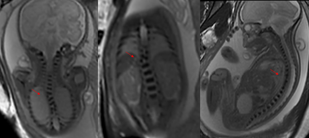
Fetal MR is often one of the best options for making a definitive diagnosis in rare cases of malpresentations or difficult ultrasound imaging. In particular, it is increasingly recognized for its value in the diagnosis of central nervous system malformations, such as neuronal migration disorders, by providing more valuable information for clinical practice. In the case of congenital anomalies of the fetal spinal cord and spine, fetal MR can further confirm ultrasound findings as an important complement.

Clinical data: 36.1 weeks of gestation; ultrasound shows a cystic structure behind the third ventricle of the fetus (arachnoid cyst to be ruled out)

Clinical data: 28.5 weeks of gestation; ultrasound shows abnormal vertebral bodies echo of the fetus' spine
Imaging diagnosis: Hypoplasia of multiple thoracic vertebrae with scoliosis
Case source: Jiedaokou Hospital and Optics Valley Hospital of Maternity and Child Healthcare Hospital, Hubei
Centering on this major medical issue, United Imaging has worked with many departments of Maternity and Child Healthcare Hospital, Hubei to promote the precise prevention and treatment of birth defects, make innovations in fetal early screening technology, popularize new modes of early screening and study fetal development and anomalies, jointly promoting medical innovation & exploration and clinical application throughout pregnancy.


