16 cm Z-axis Detector Coverage
Through-silicon-via (TSV) Technology for ultra-low noise.
3D printing technology for effective scattered photon rejection.
Cone beam artifacts mitigation associated with wide coverage systems.
Attainable Intelligence.
Simply Masterful.
As an ultra-premium 640-slice CT scanner, uCT 960+ features 16 cm z-axis detector coverage, 0.25 s rotation speed, an ultra-wide 82 cm bore, and a 318 kg. table weight capacity. Using industry-leading AI-empowered technologies, it launches the era of intelligent imaging, offering precise imaging and ease of use throughout the entire clinical spectrum.
16 cm wide Z-detector
0.5 mm detector acquisition
0.25 s rotation speed
uAI Vision
uAI CadioCapture
uAI Dose Modulation
82 cm Ultra-wide Bore
318 kg. Table Weight Capacity
60 kVp Capability
Through-silicon-via (TSV) Technology for ultra-low noise.
3D printing technology for effective scattered photon rejection.
Cone beam artifacts mitigation associated with wide coverage systems.
To deliver superior temporal resolution, freeze the motion in cardiac imaging, and enable 0.25 second rotation speed, we have a one-piece casted gantry structure integrated with a direct drive motor system providing precise rotation speed control.
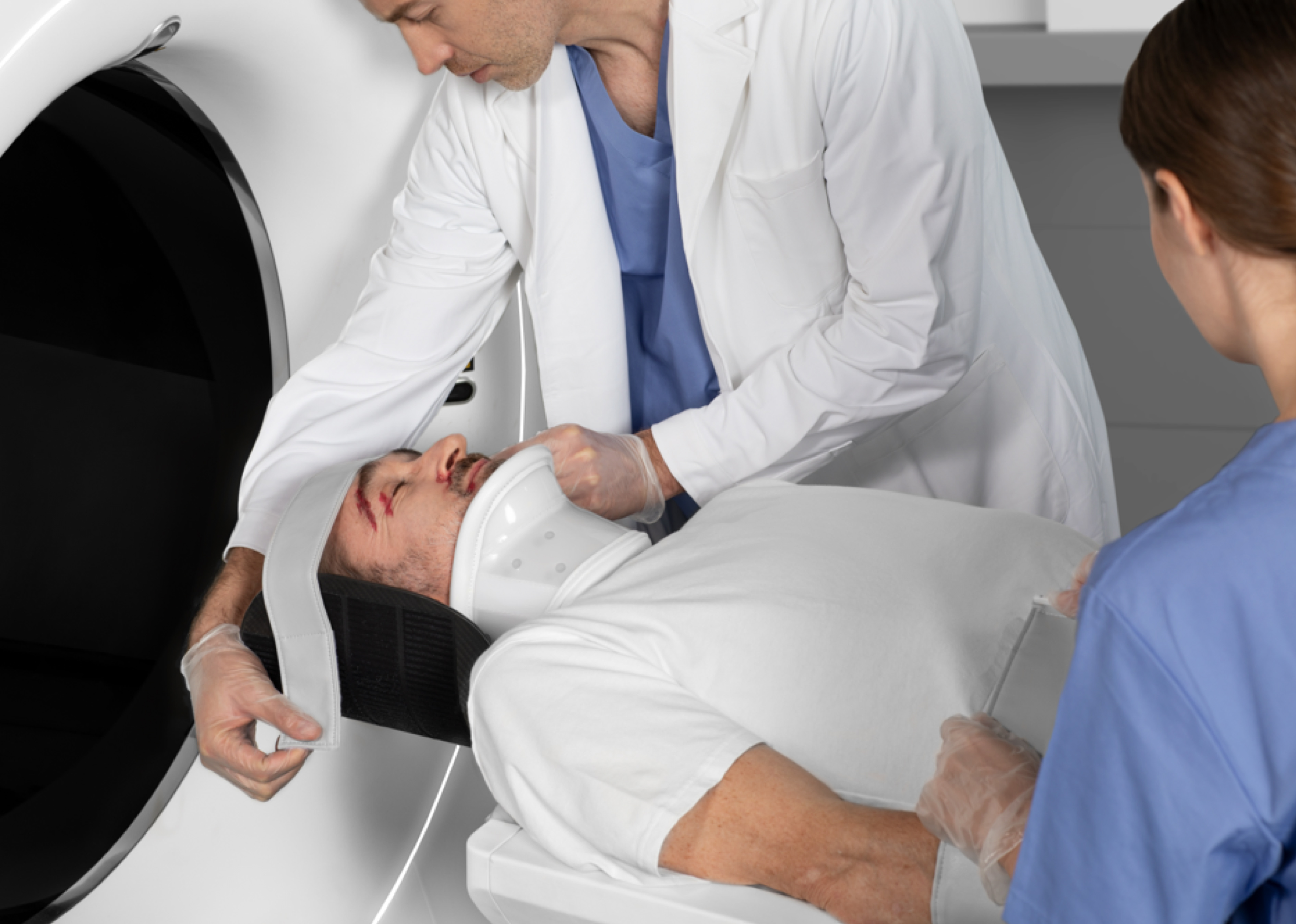
Single-click patient positioning with precise scan range locations based on the selected protocol.
Image quality is optimized taking into account the patient's surface dose distribution for every exam.
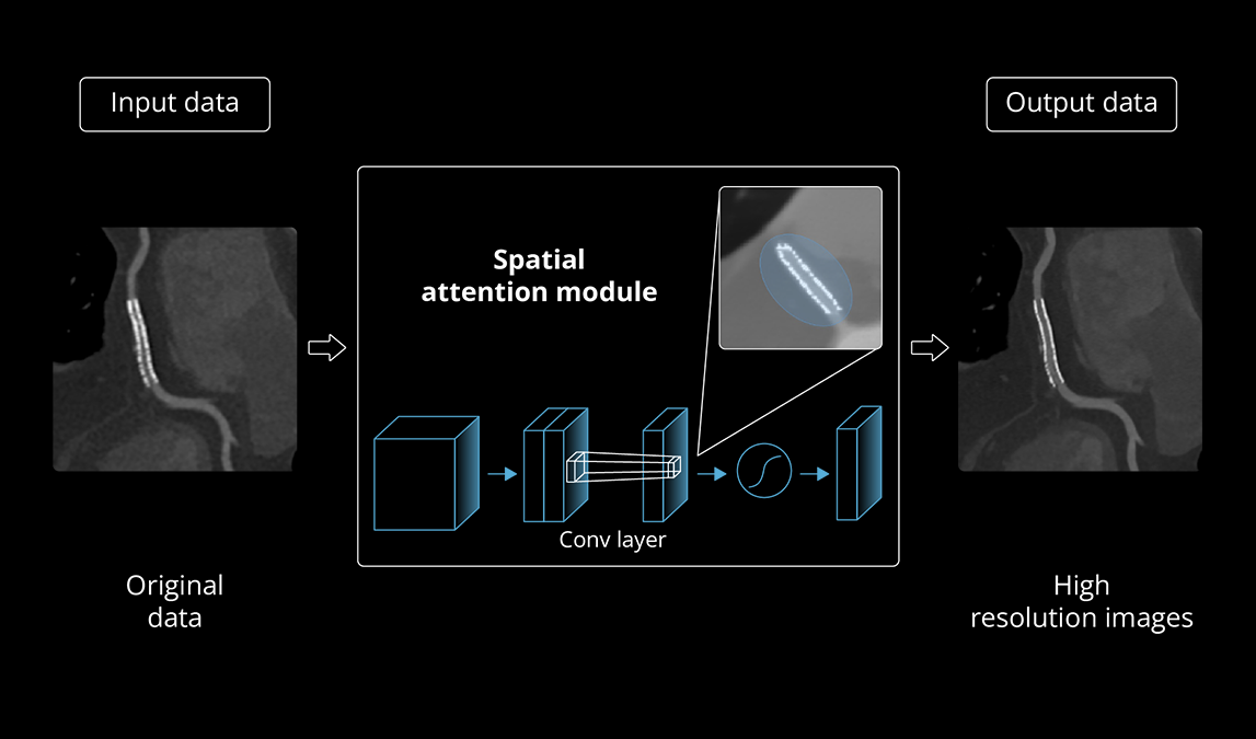
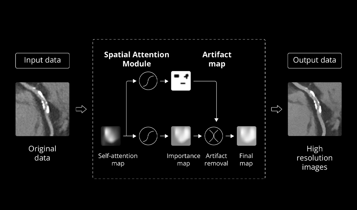
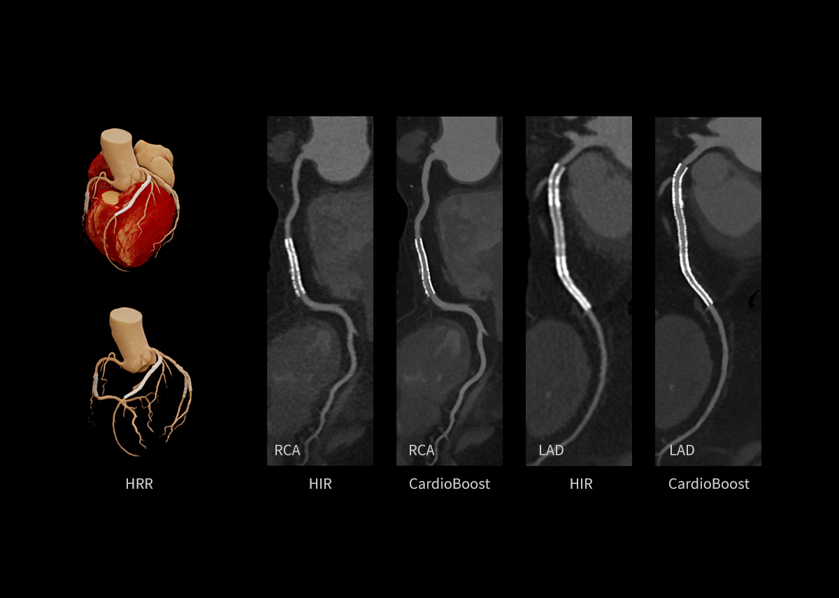
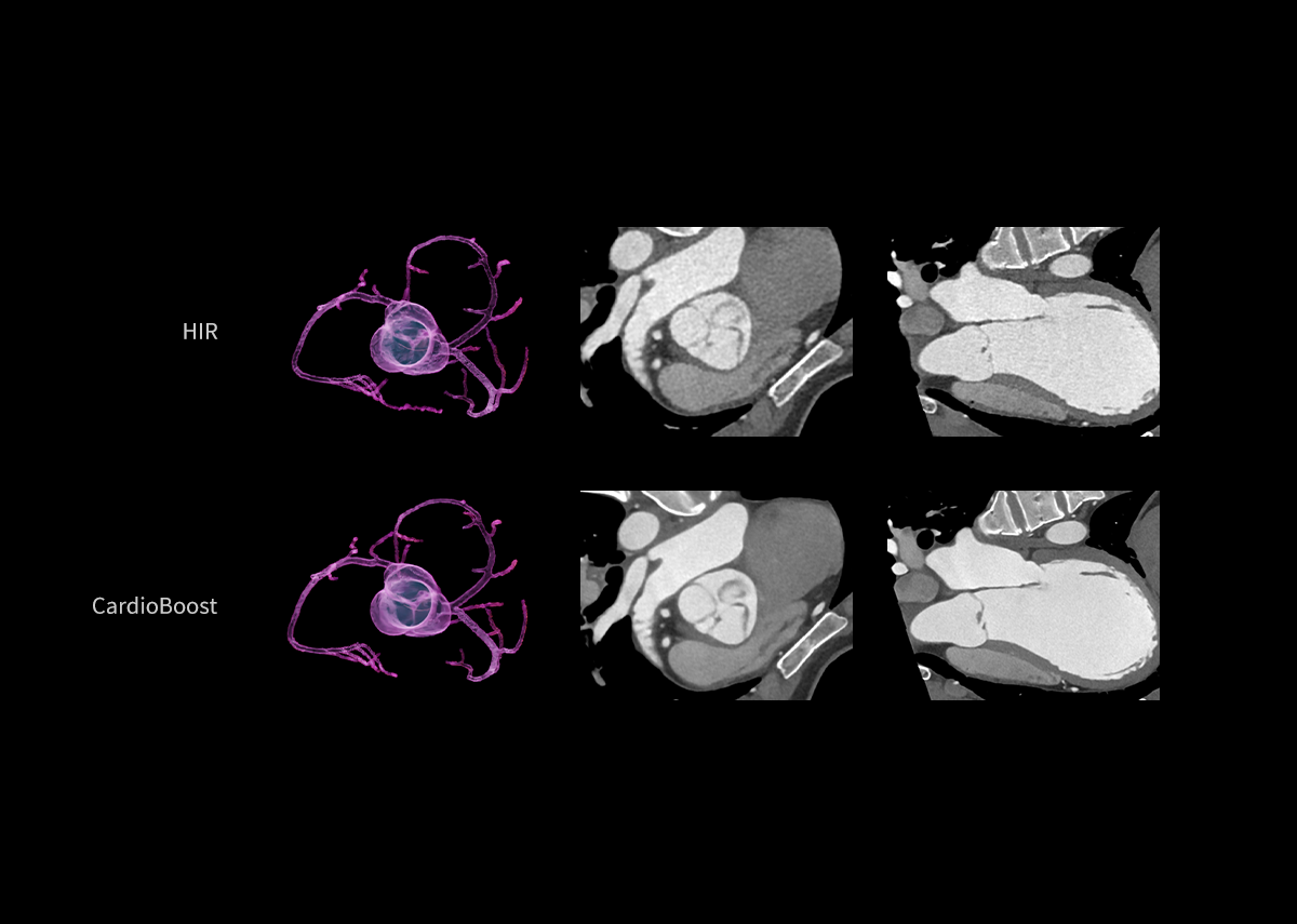
Using an intelligent motion correction with an AI-empowered coronary artery algorithm, we have improved the temporal resolution to 25 milliseconds.
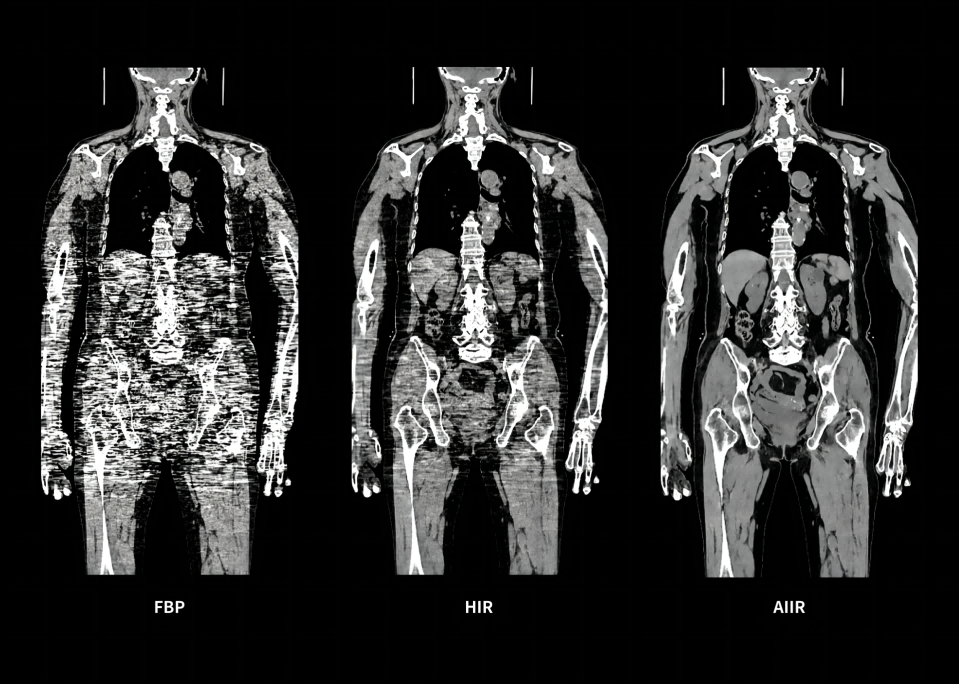
Only one target noise level resulting in under-exposing the abdomen or over-exposing the chest.
Automatic recognition of the chest and abdomen with an AI-based technology allowing a more optimal noise and optimized dose for the entire patient.
Robust, low dose and high quality cardiac imaging within a single heart beat.
CardioAssist,
CardioAdapt,
CardioXphase and
CardioCapture.
A mixed acquisition of ECG-gated axial scans and non-ECG-gated axial or helical imaging.
Quantitative and functional measurement with the ability for every scan to be spectral.
Whole organ perfusion without table motion.
Dynamic perfusion scans with large z-coverage (up to 40 cm) using a variable pitch helix.
An entire scan can be completed in half a second to minimize the motion artifacts.
One-stop evaluation of coronary arteries, pulmonary arteries, and the aorta in a single exam.
MPR or VR preview images in real-time during the acquisition.
Comprehensive and accurate intelligent rib fracture analysis.
60 kVp scans reduce radiation dose while maintaining image quality especially for smaller adults and pediatric patients.
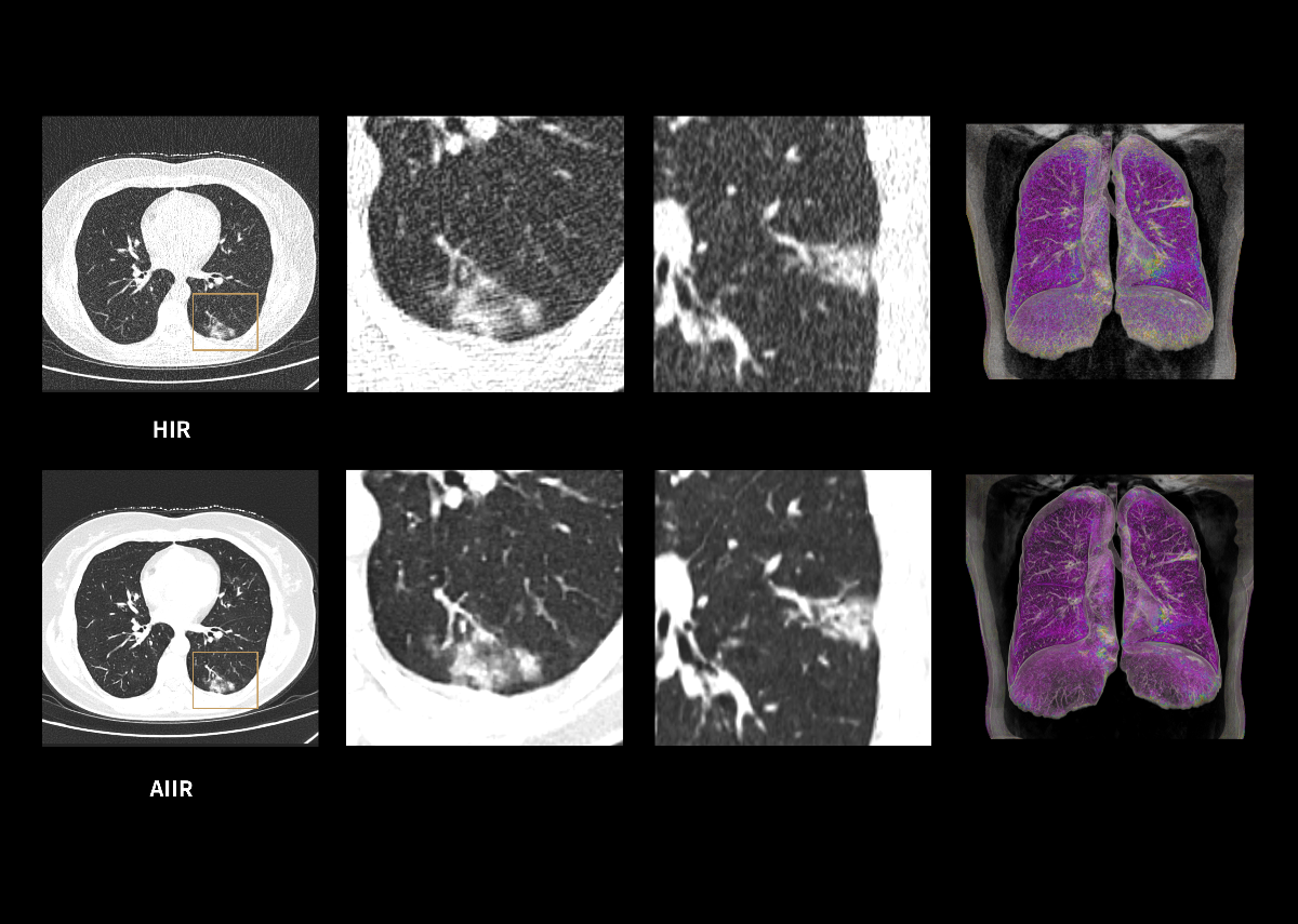
A powerful platform and AI-empowered workflow, a large daily throughput while maintaining high image quality.