
uCS® 1.5T MR
uCS® 1.5T MR
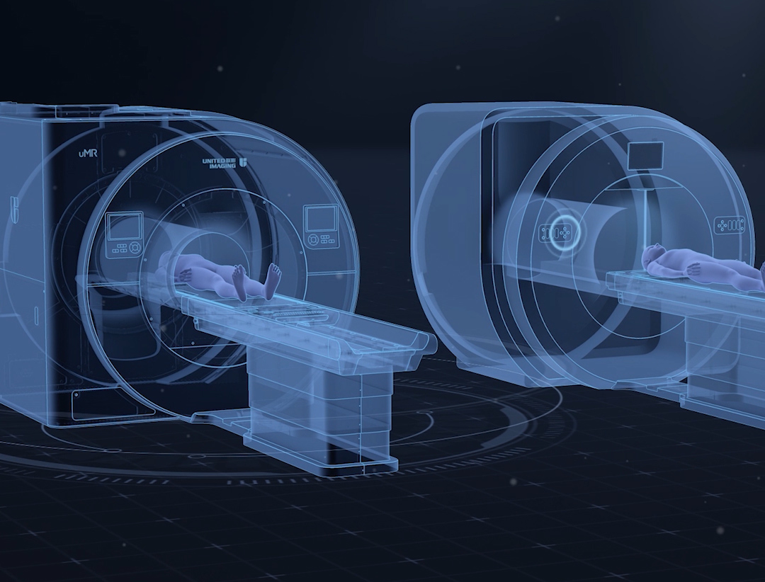
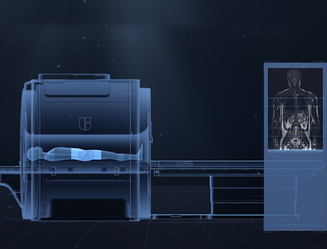
For the first time in the 1.5T field, uMR® 588 is equipped with uCS® imaging platform, which enables whole-body high speed imaging, 2s/phase abdominal dynamic multi-phase enhancement and high-resolution diffusion imaging, leading 1.5T into a new era of whole-body compressed sensing.
United Compressed Sensing
Precise Quantification
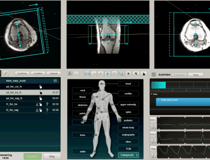
Intelligent Examination

uCS® Imaging Technology
Combining the strengths of PF, PI and CS, the uCS® (United Compressed Sensing) imaging technology has achieved optimized data acquisition and image reconstruction, significantly improving the scanning speed and image quality.
uCS® Imaging Applications
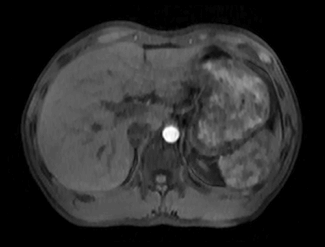
uCS® imaging is applied to the MRI abdominal dynamic scanning enhancement, which can achieve 16× acquisition acceleration and clearly capture the continuous dynamic changes of tissue signals.
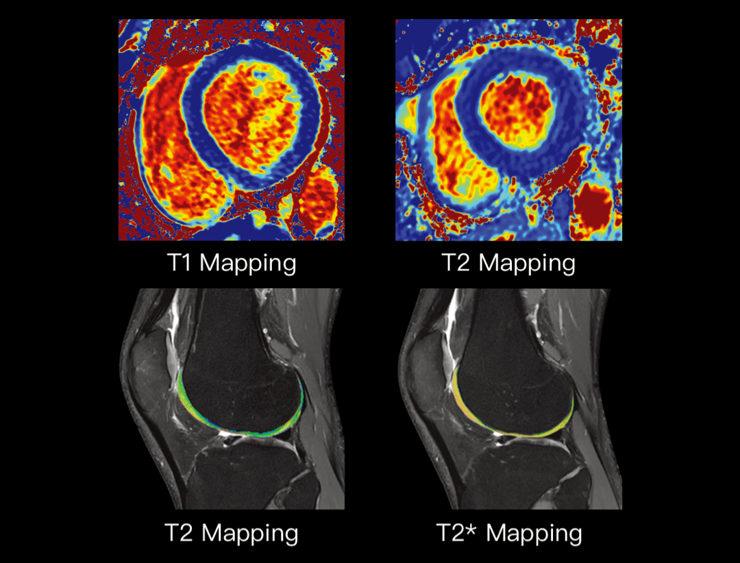
MAPs
MAPs can provide the quantitative data required for clinical and scientific research for various organ diseases by accurately calculating the time parameter.

FACT
Non-invasive detection of fat content and iron deposition in tissues. Multi-parameter images can be obtained after acquisition and calculation.
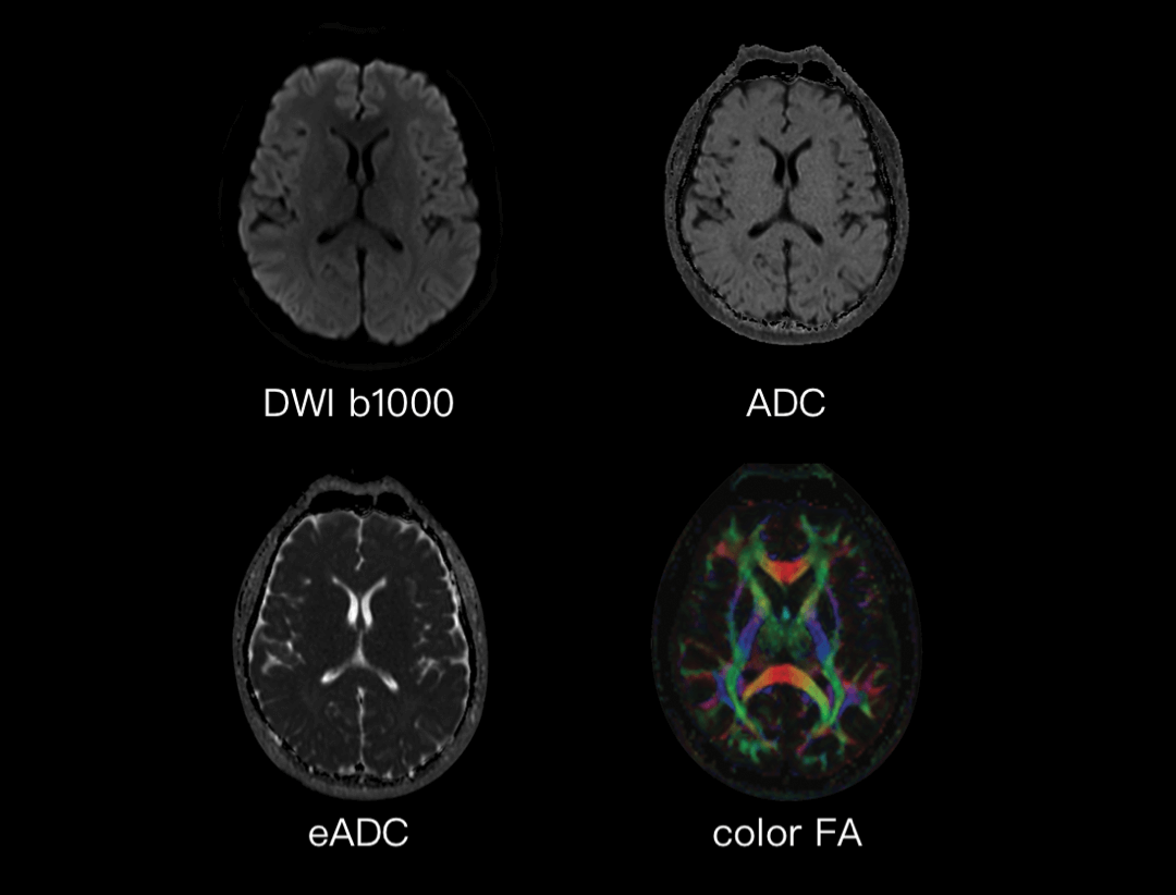
Multi-parameter Neuro Imaging
The following parameter maps can be obtained after acquisition: FA, RA, ADC, and color FA, which can reflect the speed and direction of water movement in tissue. Multi-parameter neuro imaging can provide accurate information for disease diagnosis, such as white matter lesions.
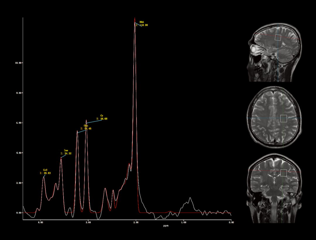
Spectroscopy
Studying the analysis of metabolic content in the human body, spectroscopy can complete the non-invasive detection of metabolite levels in living tissues.