Farsighted Exploration
Critical Illness
UC Davis' new research shows how total-body PET imaging can assess the immunological response to COVID-19 infections
UC Davis' new research shows how total-body PET imaging can assess the immunological response to COVID-19 infections
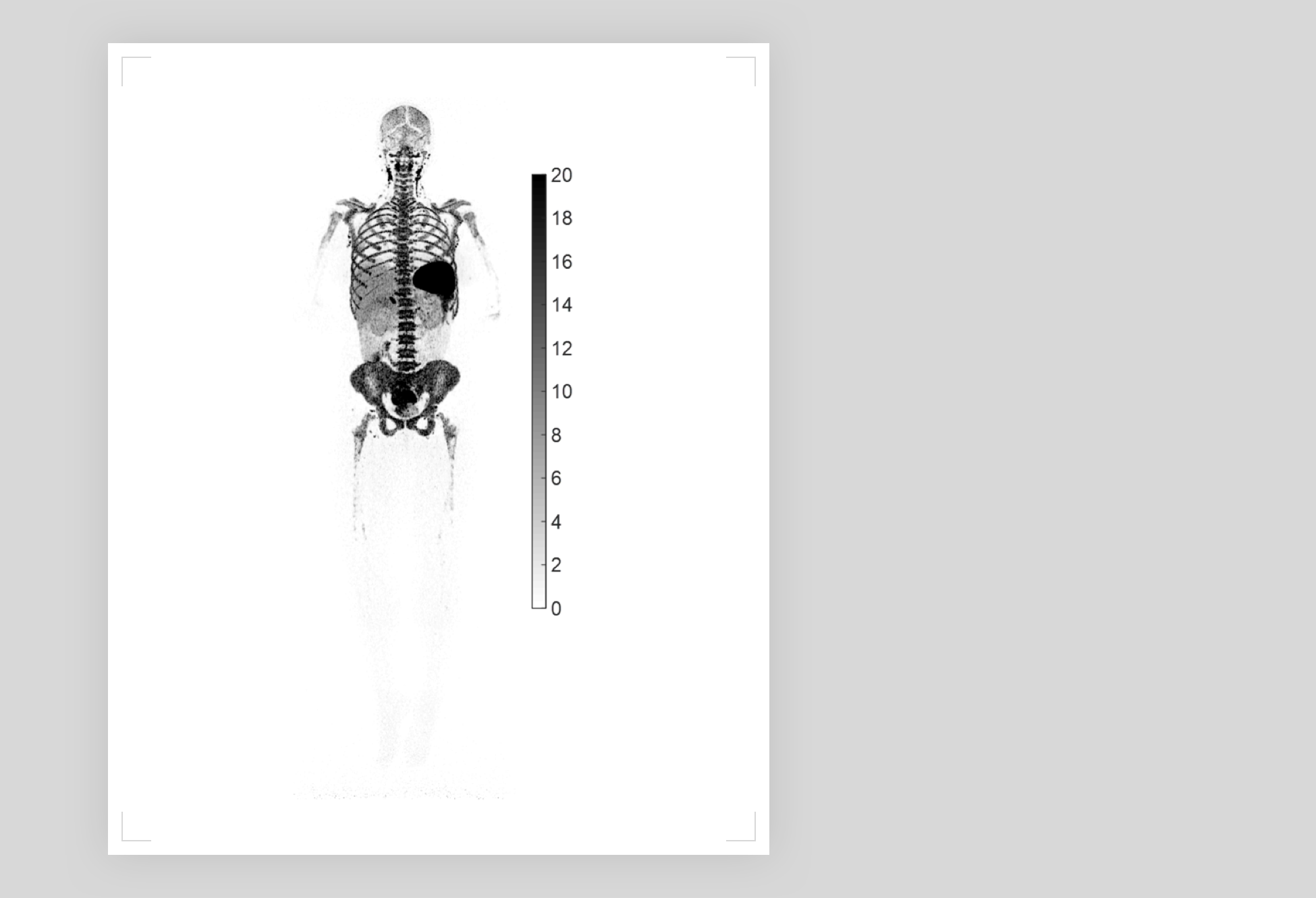
UC Davis, a leading university in molecular imaging that collaborated with United Imaging to develop the world's first total-body PET/CT scanner, 'uEXPLORER®', recently presented their research on COVID-19 at the 2022 Society of Nuclear Medicine and Molecular Imaging (SNMMI) annual meeting. Guided by the uEXPLORER scanner along with a new labeled radiotracer with high affinity to T cells (89Zr-Crefmirlimab-Berdoxam), UC Davis Project Scientist and lead author Dr. Negar Omidvari unveiled the first high-resolution PET image of total-body CD8+ T-cell distribution after COVID-19 infection. The image could shed light on the pathological mechanism and immune response to COVID-19 infection.
Sep 09, 2022
Sep 09, 2022