What structures can be imaged by bone CT scans?
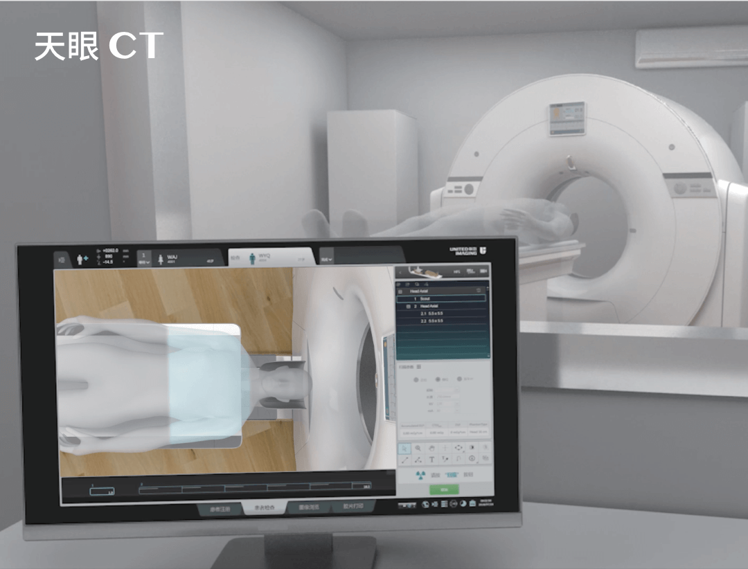
Computed tomography of the bones allows high-resolution imaging of bone structures, joints and surrounding soft tissues. Modern CT scanners, such as UIH systems, use X-rays and dedicated software to create 3D images, which allows accurate assessment of degenerative changes and injuries as well as detecting the presence of inflammatory or cancerous infiltration.
What are the indications for a bone CT scan?
Bone CT is indicated in cases where accurate assessment of bone and joint structure is required. It is usually performed in the following cases:
- injuries and fractures, especially those that are difficult to identify on ordinary X-rays;
- degenerative and rheumatic diseases where CT can reveal the extent of damage to bone structures;
- bone tumours and bone metastases where CT helps determine the size and location of lesions;
- inflammatory conditions of bones, marrow or joints, such as osteomyelitis where CT allows assessment of the extent of the inflammatory process;
- bone anatomy assessment prior to orthopaedic or endoprosthetic surgery.
Indications for bone CT also include any pain of unclear origin as well as congenital malformations and suspected cancer. Moreover, bone CT is recommended when the results of other diagnostic methods (e.g., X-ray, MRI) are inconclusive and do not allow a definitive diagnosis to be made.
SEE ALSO: CT SCAN OF THE JOINTS
Can bone CT detect cancer?
Bone CT is very often used to identify tumours and metastases in bone structures. This is because computed tomography enables precise detection of cancerous lesions (both primary tumours and secondary metastases), making it an extremely important diagnostic tool in oncology.
What can a bone CT scan detect?
Bone CT detects many different types of pathologies within the bone tissues and surrounding soft structures.
Bone CT can effectively diagnose:
- osteoarthritis;
- post-traumatic lesions, including fractures and cracks;
- dysplasias and other congenital defects;
- metabolic bone diseases, such as Paget’s disease;
- tumour infiltration and bone metastases;
- inflammatory conditions of the bone, such as osteomyelitis.
Is bone CT conducted with or without contrast enhancement?
Bone CT can be performed both with and without a contrast agent. Intravenous contrast is usually used when a more accurate assessment of the blood vessels and soft tissue surrounding the bones is required. Non-contrast examination focuses on the bone structure itself. Thus, where a CT scan of the bones is conducted without the need to analyse the surrounding tissues and blood vessels, no contrast needs to be administered.
THIS MAY ALSO INTEREST YOU: SPINE CT SCAN
Medical referral and preparation for bone CT examination
For a bone CT scan, a referral must be obtained from a doctor who will assess whether the examination is necessary based on the clinical picture. The referral should include information about the purpose of the examination and any previous imaging studies conducted. Preparation for a non-enhanced CT bone scan primarily involves the removal of metal objects, such as jewellery, which may affect image quality.
Types of bone CT examination
Bone computed tomography usually focuses on a specific anatomical area. In this regard, bone CT can be divided primarily into:
- Temporal bone CT – this examination is crucial in diagnosing inner and middle ear disorders, such as inflammation and tumours, and assessing the patient’s condition following trauma. It allows precise evaluation of temporal bone structures, which is essential when planning ear surgery or treating hearing disorders.
- CT scan of the lower leg – this examination is mainly used in the evaluation of fractures, especially those with complex morphology where standard X-ray may not provide sufficient information. It also allows assessment of the soft structures around the tibia and fibula.
- Computed tomography of the pelvic bones – this examination is extremely useful in the diagnosis of trauma, bone tumours and in assessing the patient’s condition after implantation of endoprostheses. It is also an important tool in the diagnosis of osteoarthritis of the hip.
- CT scan of the wrist – this examination is important in diagnosing wrist injuries, especially fractures of the scaphoid bone, which are difficult to detect with ordinary X-ray. It also enables the soft structures and joints of the wrist to be assessed.
Bone CT is an indispensable diagnostic tool that enables accurate imaging of bone structure, and as a result contributes to making the right diagnosis and planning effective treatment. Although the examination involves exposure to X-rays, its benefits far outweigh the potential risks in many cases. Importantly, UIH’s state-of-the-art CT scanners produce extremely precise imaging results with significantly reduced radiation doses, making bone CT even safer.
*ATTENTION! The information contained in this article is for informational purposes and is not a substitute for professional medical advice. Each case should be evaluated individually by a doctor. Consult with him or her before making any health decisions.