Spinal CT scan
What does a CT scan of the spine look like?
Computed tomography of the spine is a modern diagnostic technique that allows precise imaging of the spine and surrounding structures. Like all CT scans, computed tomography of the spine uses X-rays to create cross-sectional images. With United Imaging Healthcare’s CT scanners, the diagnostic process is supported by state-of-the-art artificial intelligence algorithms, making the examination faster, safer and even more accurate.
Unlike a classic X-ray, computed tomography provides images in multiple planes, allowing a more accurate assessment of the condition of the spine, including vertebrae, intervertebral discs and nerve structures. High-resolution images allow doctors to accurately diagnose a range of conditions from degenerative changes to trauma and cancer.
Course of the examination
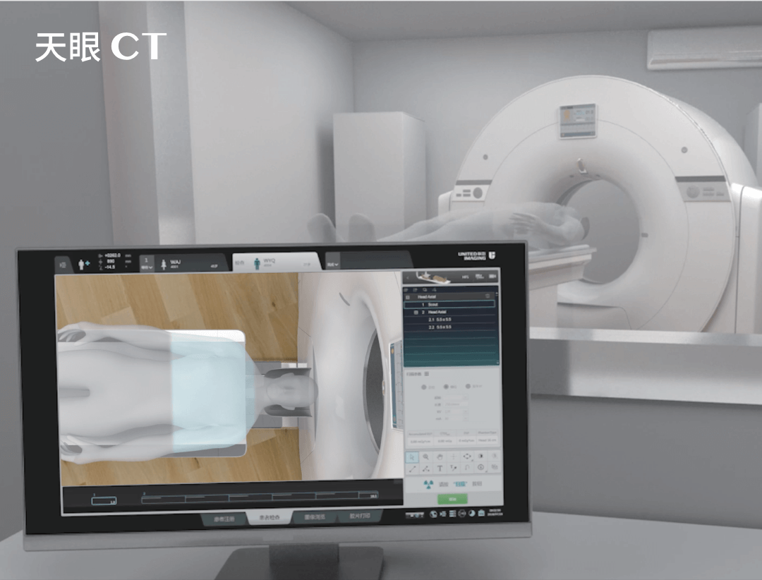
During a CT scan of the spine, the patient lies on a special table that slides inside a bore of the CT scanner, which is equipped with detectors and X-ray tubes. The tubes emit a beam of rays that passes through the patient’s body and is subsequently recorded by detectors. This information is transmitted to a computer, which processes it, creating a two- or three-dimensional image of the body part examined.
What can a CT scan of the spine show?
CT scans make it possible to detect many serious spinal conditions at an early stage. This examination makes it possible to diagnose the following conditions:
- degenerative changes of the spine, including degeneration of intervertebral discs and calcifications;
- herniated intervertebral discs, which may press on the spinal cord or nerve roots;
- spinal injuries such as compression fractures, damage to intervertebral joints and ligaments;
- cancer, including primary tumours of the spine and metastases;
- inflammatory and infectious conditions, such as ankylosing spondylitis;
- spine damage caused by accidents and injuries.
What diseases can a CT scan of the spine detect?
Computed tomography of the spine is useful in diagnosing a wide variety of pathological conditions, most notably:
- osteoporosis and its consequences, such as pathological fractures;
- spondyloarthropathies, including Bechterew’s disease (ankylosing spondylitis);
- congenital anomalies of the spine, such as scoliosis or spina bifida;
- spinal pain of unclear origin.
THIS MAY ALSO INTEREST YOU: CARDIAC CT
Is it necessary to be fasted for a spinal CT scan?
Typically, a patient does not need to be fasted before a standard spinal CT scan, unless contrast agent administration is envisaged. In this case, the patient may be required to fast for several hours before the examination. Detailed preparation guidelines should always be provided by the facility performing the scan.
How to prepare for a CT scan of the spine?
As mentioned above, preparation for spinal CT scans may require fasting if a contrast agent will be used. The patient should also inform the doctor about any medications he or she is taking and about any allergies, especially to contrast agents. In addition, it is important that all metal objects, such as jewellery, which may interfere with image quality, be removed before the examination.
Is a CT scan of the spine safe?
Computed tomography of the spine is a safe examination, although it uses X-rays, which may raise concerns about radiation exposure. However, the diagnostic benefits of accurate imaging usually far outweigh the risks. However, CT scan of the spine should be avoided in pregnant women, especially in the first trimester, due to potential risks to the foetus.
THIS MAY ALSO INTEREST YOU: CARDIAC MRI
Contraindications to CT scans
In addition to pregnancy, a CT scan may be contraindicated if the patient is unable to remain motionless during the examination. The cause may be nervous tics or Parkinson’s disease. Unfortunately, any patient movement during the scan can lead to inaccurate and inconclusive results. CT scans are also contraindicated in claustrophobia. Patients who suffer from claustrophobia, as well as those who are hyperactive, may be administered sedatives to calm and soothe them.
Which is better: an MRI or a CT scan of the spine?
The choice between magnetic resonance imaging (MRI) and computed tomography (CT) depends on the type of condition being diagnosed and the specific diagnostic requirements. MRI is usually the method of choice for examining soft tissues such as intervertebral discs, ligaments and the spinal cord. CT, on the other hand, is indicated in the evaluation of bone structures, trauma, and in cases where MRI is contraindicated (such as patients with metal implants). The final choice of imaging modality should always be made by the attending physician based on an individual assessment of the patient’s condition.
Computed tomography of the spine is an invaluable tool in the diagnosis of many serious spinal conditions, offering detailed images that enable accurate diagnosis and effective treatment planning. Its ability to detect a wide spectrum of pathologies makes it the keystone of modern diagnosis of spinal conditions.
*ATTENTION! The information contained in this article is for informational purposes and is not a substitute for professional medical advice. Each case should be evaluated individually by a doctor. Consult with him or her before making any health decisions.
SEE ALSO: WHAT IS CT PERFUSION?