Cardiac MRI – indications, what does it look like, how to prepare for the scan and how long does a MRI scan take?
Cardiac MRI is a medical imaging procedure that uses a strong magnetic field and radio waves to obtain detailed images of the heart’s structure and function. This technology enables diagnostic assessments of the myocardium and valves as well as blood vessels with an accuracy that cannot be achieved using other methods. This examination is also often used to complement echocardiography and CT scans, providing more accurate information about the heart’s condition.
What does the examination consist in?
Cardiac MRI leverages the magnetic properties of the particles present in atomic nuclei, mainly protons. Under normal conditions, these protons have different spin orientations. During a magnetic resonance imaging (MRI) scan, the patient is placed in a strong magnetic field that orients the protons’ spins in a single direction. Subsequently, electromagnetic pulses are used to excite atomic nuclei, imparting additional energy to them. Once the pulses are over, the atoms begin to release the stored energy, which occurs in two ways, known as T1 and T2 relaxation. The energy thus released is recorded by the coils as a change in voltage, and the data are converted into images by a computer.
In some cases, it is necessary to administer a contrast agent containing gadolinium to the patient. It is administered through an intravenous line. In the images obtained, the inflamed area appears brighter compared to healthy myocardial tissue, which quickly flushes out the contrast agent. This is called late gadolinium enhancement (LGE). In addition, pericardial effusion is also monitored during the examination.
SEE ALSO: COMPUTED TOMOGRAPHY OF THE HEART
When should a cardiac MRI be performed?
The examination is recommended in a variety of clinical situations where cardiac pathology is suspected or cardiac structure or function needs to be accurately assessed. A huge advantage of a cardiac MRI scan is its high accuracy and relatively low burden on the patient. Some of the main indications for cardiac MRI include:
- Myocarditis: cardiac MRI can be performed to diagnose and monitor myocarditis caused by an infection or autoimmune disease.
- Coronary artery disease: cardiac MRI can be used to evaluate blood flow through the coronary arteries and identify areas of myocardial ischemia or scarring.
- Cardiomyopathies: the examination is recommended in the diagnosis of various types of cardiomyopathies, including dilated, hypertrophic and restrictive cardiomyopathy, since it enables the assessment of the structure, function and thickness of the myocardium.
- Heart defects: the examination can help diagnose valvular defects, myocardial wall defects or abnormalities in the structure of blood vessels.
- Diagnosis of cardiac tumours: if the presence of tumours, including cancer, within the heart is suspected, this examination can help identify, localise and stage them.
- Follow-up after cardiac procedures: following heart surgeries such as stent, bypass or pacemaker implantation, cardiac MRI may provide a key method for monitoring the progress of treatment and assessing possible complications.
In each case, however, it is necessary to consult a doctor who will assess whether an MRI scan is indicated in a given clinical situation. The timing of the examination will depend on the urgency of the diagnosis and the treatment plan set by the specialist.
Contraindications to the examination
Although cardiac MRI is a relatively safe diagnostic procedure, patients should carefully discuss their health situation with their doctor before undergoing the examination to assess any possible risks and contraindications. If there is any doubt, additional tests may be ordered or alternative diagnostic methods may be suggested. Among the contraindications to cardiac MRI, special attention should be paid to:
- the presence of metal implants that are not compatible with an MRI scan;
- first trimester of pregnancy;
- active or recent infection or taking antibiotics;
- cardiac arrhythmias, atrial fibrillation, and other conditions that can make ECG gating difficult;
- allergy to the contrast agent if it is to be administered.
How to prepare for the examination?
The patient should undergo the examination fasted, which means that the last meal should be eaten four to six hours before the scheduled MRI scan. Exceptions are medications that the patient takes on a permanent basis – these can be taken at normal times with a small amount of still water.
When preparing for the procedure, the following recommendations should also be followed:
- Avoid consuming coffee, tea, chocolate and energy drinks two days before the examination.
- Exercise and smoking should be avoided on the day of the examination, since these activities can increase the heart rate and chest movements, which in turn can affect the quality of the images obtained during the scan.
- On the day of the examination, avoid using cosmetics containing metallic particles, as they may interfere with the image.
- It is advisable to use the restroom before the examination, as the scan cannot be interrupted.
- If necessary, the patient’s chest should be partially shaved before the examination so that the electrodes can be attached.
Before a contrast agent is administered, it is important to assess renal function. For this purpose, a blood creatinine level test should be performed beforehand. In some cases, a glomerular filtration rate (GFR) assessment is also recommended, which is based on an analysis of a blood and urine sample.
MORE ON THIS SUBJECT: HOW TO PREPARE FOR AN MRI SCAN?
What does a cardiac MRI look like?
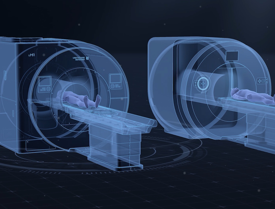
A cardiac MRI scan includes several stages. At the outset, the patient may be asked to remove any metal objects that may interfere with the magnetic field (this may include watches, jewellery or other metal objects). The patient is then placed on an examination table, which moves inside a cylindrical bore of the MRI machine. It is very important that the patient’s entire chest be inside the machine. Electrodes, similar as those used in an ECG examination, are glued to the patient’s chest. This allows the patient’s heartbeat to be accurately monitored and the scanner to be properly synchronised with it.
Throughout the examination, medical staff monitor the patient’s condition to ensure safety and comfort. The patient is also in constant contact with staff using the intercom built into the MRI machine.
Once a series of images has been acquired, the patient is released and the examination is completed. The results are then passed on to the doctor who interprets them and continues diagnostic procedures or initiates treatment.
MRI examination is painless and usually takes between 30 and 60 minutes, but its duration may vary depending on the complexity of the case and diagnostic requirements. During the procedure, it is important that the patient remain motionless. Some discomfort may be caused by the relatively high noise level generated by the machine, but this may be reduced to some extent by headphones worn by the patient. The machine’s narrow bore can also induce claustrophobia. For this reason, young children are most often sedated before the examination.
Cardiac MRI is an indispensable diagnostic tool enabling precise assessment of the heart’s structure and function. With this examination, it is possible to make a quick diagnosis and choose the optimal treatment strategy for the patient. Therefore, the patient should prepare well for the examination and consult a specialist about possible contraindications to avoid unnecessary complications and guarantee the safety of the entire procedure.
*ATTENTION! The information contained in this article is for informational purposes and is not a substitute for professional medical advice. Each case should be evaluated individually by a doctor. Consult with him or her before making any health decisions.
THIS MAY ALSO INTEREST YOU: WHAT IS CT ANGIOGRAPHY?


