Prostate MRI scan – indications, scan duration and contraindications
This non-invasive method makes it possible to obtain detailed 3D images, enabling doctors to accurately assess the condition of the prostate, which is invaluable for precise treatment planning.
Indications for a prostate MRI scan
Magnetic resonance imaging (MRI) of the prostate plays a key role in advanced urologic diagnosis, especially when elevated prostate-specific antigen (PSA) levels or concerning findings on per rectum examination (rectal palpation) indicate the possibility of cancer.
Trivia: the PSA test is a commonly used screening tool that can indicate an increased risk of prostate cancer, but it is not sufficiently specific to confirm a diagnosis. MRI of the prostate allows for a much more accurate assessment, providing high-resolution images that enable even small changes in prostate tissue to be identified.
One of the main advantages of MRI is its ability to differentiate between tumours confined to the prostate and those that have spread beyond the gland itself. The test also helps assess the invasion of cancer into adjacent structures, such as the bladder or lymph nodes, which is important for correctly staging the disease. Owing to MRI imaging, doctors can also pinpoint which areas of the gland are most suspicious and must be biopsied, making prostate cancer diagnostics more effective.
MRI is also an invaluable tool in assessing response to treatment, such as after radiotherapy or surgery. Monitoring changes in prostate tissue using MRI enables early detection of recurrence or assessment whether treatment has achieved the intended therapeutic effect.
SEE ALSO: CT UROGRAPHY – WHAT IS URINARY TRACT TOMOGRAPHY?
Contraindications to the examination
Safety is a top priority, so the patient should discuss any potential risk factors in detail with the doctor prior to the examination. Particular challenges during the examination may be posed by:
- metal implants;
- a pacemaker;
- vascular stents or clips;
These can not only interfere with the quality of MRI images, but in some cases they can also pose a risk to patient safety, for instance by heating up or moving under the strong magnetic field generated by the MRI machine.
Another aspect which should be carefully considered is a potential allergy to the contrast agent used in some MRI examinations. Although allergic reactions to contrast agents are rare, their consequences can be serious, and thus it is important to discuss any known allergies with the doctor before the examination, especially if the patient has had previous reactions to contrast agents used in imaging studies. If the risk of an allergic reaction has been identified, the doctor may decide to administer a different contrast agent, administer allergy medication or forgo the use of contrast.
How to prepare for a scan?
Preparing for the examination usually does not require changes in daily routine. The procedure is a little different, however, when the administration of a contrast agent is envisaged.
In this case, the patient may be advised to discontinue certain medications to reduce the risk of potential interactions with the contrast agent. In addition, blood tests may be required to ensure that the patient’s kidneys are functioning properly and are able to safely excrete the contrast agent.
It is also recommended that the patient be fasted before the examination. This is a precautionary measure to minimise the risk of nausea or vomiting, which may occur in a small number of patients.
Patient comfort during the examination is crucial as well, and thus it is advisable to dress in comfortable clothing that enables free movement. The clothing worn should be free of metal parts such as buttons, zippers or buckles that could interfere with image quality. Patients should also remove jewellery and other metal objects that may affect the examination.
MORE ON THIS SUBJECT: HOW TO PREPARE FOR AN MRI SCAN?
Course of the examination
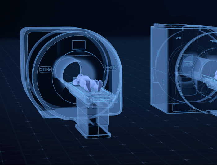
During the examination, the patient lies supine on a moving table that slides inside the bore of the MRI machine. The examination usually lasts between 30 and 60 minutes, and the patient must remain motionless in order to ensure high image quality. MRI is completely painless, although the sounds generated by the machine can be loud, and therefore patients are often offered headphones or earplugs.
The patient can usually return to normal activities directly after the examination. If a contrast agent is administered during the examination, it is recommended to increase fluid intake to help excrete it from the body.
#BBD0E0 »


