Pelvic MRI: applications, course of examination, contraindications
Magnetic resonance imaging (MRI) of the pelvis is an advanced imaging test that can be used to diagnose diseases of the genitourinary system, including conditions of the bladder, prostate, ovaries and uterus as well as joint and muscle structures of the pelvis. It is an indispensable tool in detecting tumours, cysts, inflammation and endometriosis as well as in assessing the integrity of anatomical structures and detecting possible congenital anomalies.
This examination is considered one of the safest diagnostic methods available to modern medicine due to the fact that the patient is not exposed to ionising radiation. This is the difference between MRI and other popular imaging techniques such as computed tomography (CT) or traditional X-rays, which, despite their diagnostic value, carry certain risks.
Owing to the fact that MRI machines include advanced software, accurate images of the body’s internal structures can be obtained, which makes this method particularly attractive to patients and doctors.
Indications for pelvic MRI scan
One of the main indications for this examination is where a tumour is suspected. Pelvic MRI allows not only to identify the presence of tumours in the patient’s body, but also to accurately determine their size, location and potential spread, which is essential for planning an appropriate treatment strategy.
In addition, MRI allows the following conditions to be diagnosed:
- inflammatory conditions including evaluation of their severity as well as identification of potential complications such as abscesses or fistulas;
- endometriosis, which is a chronic disease affecting women and involves the presence of tissue similar to the endometrium outside the uterine cavity.
Magnetic resonance imaging is invaluable in assessing the integrity of anatomical structures of the pelvis, allowing possible anomalies that may affect urogenital function and other vital functions to be detected. Their early detection is key to planning appropriate medical or surgical intervention.
Preparation for the examination
Before undergoing an MRI of the pelvic area, it is recommended that the patient be fasted for at least six to eight hours before the scheduled examination, and take care to empty the bowels. On the day before the examination, the patient may also be asked to take a laxative as prescribed by the doctor, as well as an agent reducing intestinal gas to minimise bloating.
In addition, the patient is advised to wear loose, comfortable clothing without metal parts that could interfere with the magnetic field.
MORE ON THIS SUBJECT: HOW TO PREPARE FOR AN MRI SCAN?
Contraindications to pelvic MRI
Although the examination itself is a safe and effective diagnostic method, there are certain contraindications to its performance. Patients with metal endoprostheses must provide accurate information to medical staff about the material from which these implants are made in order to assess whether the examination can be performed safely.
For patients with pacemakers, other electrical implants such as neurostimulators, or insulin pumps, conducting an MRI scan in an outpatient setting, where the patient is not hospitalised and leaves the medical facility immediately after the examination, is not possible due to the potential risk of interference with these devices.
In addition, patients who have had clips or vascular stents implanted may not undergo pelvic MRI until six to eight weeks after surgery, due to the need for the wounds to heal completely and for the implanted materials to be stabilised.
Special attention should also be paid to where MRI scans are to be conducted in pregnant women. It is recommended that MRI scans be avoided during the first trimester of pregnancy unless there are important medical indications and the test has been consulted with a gynaecologist. The decision to perform an MRI at this particular time should always involve consideration of the potential benefits and risks to the health of the mother and the foetus.
For patients suffering from claustrophobia, the examination can be attempted after taking sedatives, which must be prescribed in advance by a general practitioner or specialist, and thus prior consultation with the doctor is advised.
SEE ALSO: MRI IN PREGNANCY
Course of the examination
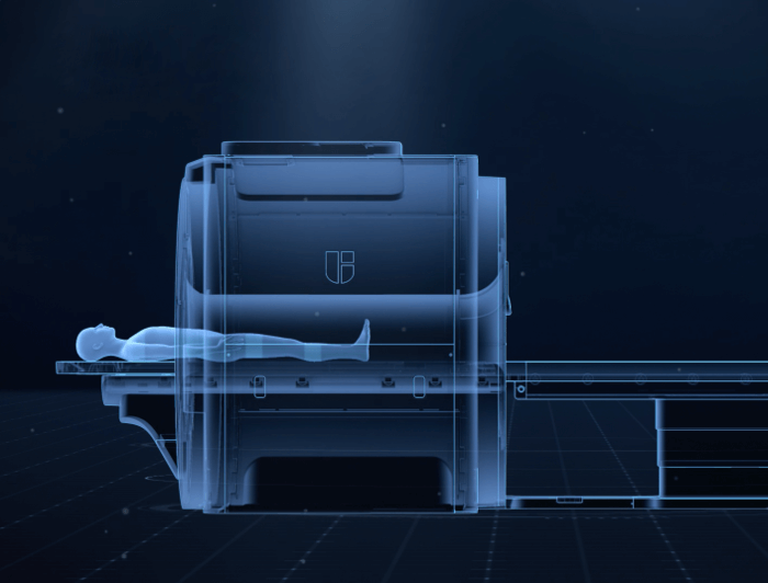
During an MRI scan, the patient lies on a moving table that slides inside the bore of the MRI machine. The machine emits short magnetic pulses that interact with hydrogen atoms in the body, allowing images of internal body structures to be obtained.
The procedure is completely painless, but requires the patient to remain motionless for the duration of the examination, which typically ranges from 40 to 70 minutes depending on the area to be examined and the image detail level required.
After the examination has been completed, the images are analysed by a radiologist who specialises in interpreting MRI results. Careful image analysis makes it possible to identify or rule out various conditions, plan further diagnostics or treatment, and monitor the progress of treatment of already diagnosed conditions.
THIS MAY ALSO INTEREST YOU: MRI VERSUS CT SCANS – THE DIFFERENCES
*ATTENTION! The information contained in this article is for informational purposes and is not a substitute for professional medical advice. Each case should be evaluated individually by a doctor. Consult with him or her before making any health decisions.


