CT colonography – what is it, who is it for and how should I prepare?
CT colonography, also known as virtual colonoscopy, is an advanced imaging test that provides a detailed assessment of the inside of the colon. Using X-rays and computer data processing, CT colonography produces 3D images of the colon, allowing its structure and function to be accurately assessed. This examination is invaluable in diagnosing various pathological conditions such as polyps, tumours, diverticula or other structural abnormalities of the colon.
How does CT colonography work?
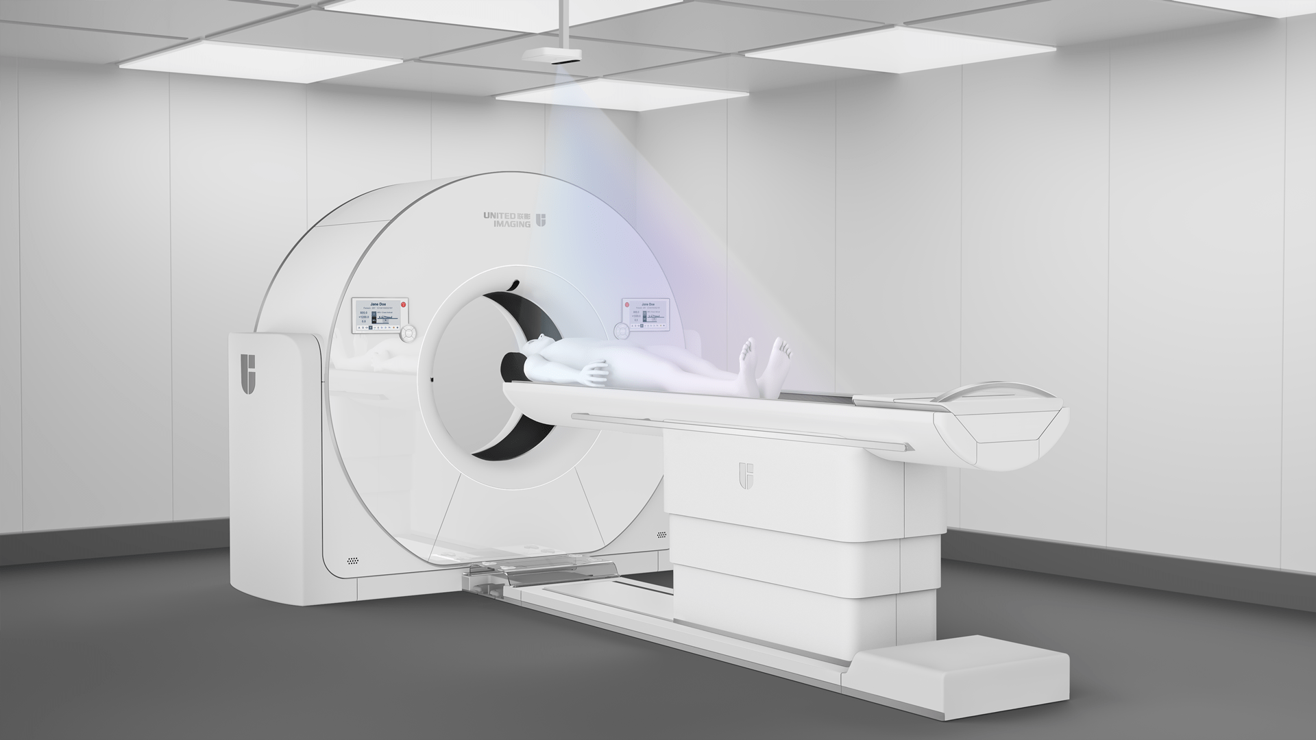
The examination is performed using a computed tomography (CT) scanner. During the procedure, the patient lies first in a supine position, and then in a prone position on a moving table, which moves through the ring-shaped gantry of the scanner, while the physician performs the imaging examination. Before imaging, the patient’s colon is gently filled with air or carbon dioxide, which makes it possible to obtain more detailed and clearer images. A computer processes the data obtained using the X-rays to create cross-sectional images (colon scans), which can then be converted into a 3D image.
The advantages of this method are that it is simple and well tolerated by patients. It allows not only colon, but also other structures within the abdominal cavity to be imaged. In addition, CT colonography is a relatively sensitive imaging method; this is, however, dependent on a thorough emptying of the colon.
Indications to the examination
Owing to its precision and detail, this examination allows lesions to be detected at an early stage, which is crucial for effective treatment and prevention of complications associated with colorectal diseases. Indications for this examination include:
- diagnosing polyps and early stages of colorectal cancer in individuals from risk groups;
- diagnosing causes of complaints such as abdominal pain, changes in the pattern of bowel movements, rectal bleeding;
- evaluation of the colon in patients who have undergone treatment for colorectal cancer to detect possible recurrence.
In addition, this examination is also performed when contraindications to a standard colonoscopy are found or the result of that examination is insufficient.
Contraindications to CT colonography
CT colonography is a safe imaging method for patients, but there are some contraindications and situations in which caution should be exercised and the patient’s health condition should be discussed with the referring physician. Prominent among these situations are:
- pregnancy;
- acute inflammation of the intestines, which may be a contraindication to filling the bowel with air;
- intestinal perforation;
- recent intestinal surgery;
- severe heart and lung diseases.
It is important to inform the doctor about any pre-existing medical conditions, past surgeries, as well as possible allergies to contrast agents before the examination. This can minimise the risk of complications and ensure that the examination is conducted safely.
How do I prepare for the examination?
Proper preparation for CT colonography is crucial in order to obtain accurate results. Two days before the colonography, the patient should switch to a low-fibre diet, which means avoiding vegetables, fruits, pasta and bread (especially multigrain bread). However, small amounts of hardtack can be consumed. Meals should be based on dairy (milk, yoghurt), eggs and cooked meat.
The day before the colonography, the patient should start taking barium suspension, which must be collected from the hospital reception at least three days in advance. It should be taken with each of the three meals (breakfast, lunch, dinner) – the patient should consume 20 ml of the suspension each time.
In the afternoon, between 4:00 and 5:00 pm, the patient should drink an Epsom salt solution (1 tablespoon dissolved in a glass of water), which is available from pharmacies without a prescription. At 7:00 pm, the patient should take 4 tablets of Bisacodyl, which is also available without a prescription.
About 2–3 hours before the examination, contrast is administered orally in a hospital setting. Only sugar-free liquids, such as still water or tea, can be consumed on this day.
After the examination
After the examination, the patient may experience slight bloating or discomfort from the introduction of air into the intestine, but these symptoms usually resolve quickly. The patient can return to normal activities as soon as he or she leaves the hospital. It is also recommended that the patient drink plenty of fluids and eat fruits to avoid constipation and help flush the contrast agent out of the body. It should also be remembered that immediately after the examination, stools may be harder and lighter in colour.
Who can order this examination?
Any doctor can make a referral for CT colonography, but procedures vary depending on whether the examination is paid for by the patient privately or funded by the National Health Fund. In the case of private payment by the patient, a referral can be made by a family doctor or specialist who will assess the need for the examination. However, for examinations reimbursed by the National Health Fund, a referral from a specialist such as a gastroenterologist, surgeon or oncologist is required.
*ATTENTION! The information contained in this article is for informational purposes and is not a substitute for professional medical advice. Each case should be evaluated individually by a doctor. Consult with him or her before making any health decisions.


