What is imaging diagnostics?
Imaging is the cornerstone of modern medicine and encompasses a range of advanced technologies used to visualise the inside of the human body. It provides unprecedented insight into the structure and function of the body and has become an essential tool in the diagnosis, treatment and monitoring of a wide range of conditions. What exactly is diagnostic imaging? What are the differences between medical imaging techniques? And what milestones in its development have contributed to significant advances in healthcare?
The basics of diagnostic imaging
Diagnostic imaging, which encompasses a range of techniques and procedures, is used to image the human body for diagnostic and therapeutic purposes. It is a rapidly developing field of medicine that allows detailed analysis of a patient’s condition. Diagnostic imaging allows the visualisation of both external and internal body structures, providing vital information that may not be available through conventional examination methods.
Diagnostic imaging uses a variety of techniques such as digital radiography (DR), ultrasound (US), computed tomography (CT) or magnetic resonance (MR). Each of these techniques has its own specific applications, advantages and disadvantages, and the choice of the appropriate method depends on the specific medical case.
Diagnostic imaging – history and breakthroughs
The origins of diagnostic imaging date back to the late 19th century when Wilhelm Conrad Röntgen discovered X-rays. In 1895, his experiments led to the first X-ray image – of his wife’s hand. This was a turning point in medicine, paving the way for new diagnostic possibilities.
In the decades that followed, imaging technology developed rapidly. A key moment was the development of computed tomography (CT) in the 1970s. Its pioneers – Godfrey Hounsfield and Allan Cormack – were awarded the Nobel Prize in 1979. CT, which combines X-rays with computer algorithms, made it possible to produce cross-sectional images of internal body structures with remarkable accuracy.
Developments and innovations in medical imaging
Another breakthrough came in the 1980s with the introduction of magnetic resonance imaging (MRI). Using powerful magnetic fields and radio waves, MRI revolutionised soft tissue diagnostics by providing high-resolution images without exposing patients to radiation. Meanwhile, innovations in ultrasound, including the development of echocardiography, made it possible to examine the heart and other internal organs more accurately in real time.
In recent years, we have seen further advances in imaging techniques, including their digitalisation and automation, which are significantly improving the quality and availability of such examinations. Importantly, United Imaging Healthcare has been at the forefront of these developments.
The development of imaging tests using radioactive substances in nuclear medicine is opening up new possibilities in the diagnosis and treatment of cancer and neurodegenerative diseases. The implementation of artificial intelligence algorithms is making it possible to speed up examinations and minimise risk factors. Each of these factors contributes to improving the accuracy, safety and efficiency of diagnostic imaging, making it an integral part of modern healthcare.
X-rays – the foundation of medical imaging
X-ray imaging, or X-ray for short, is the oldest technique in the field. It produces two-dimensional images of different parts of the body. These images show differences in tissue density and can be used to detect problems such as broken bones, lung infections or even cancer. Modern X-ray techniques, including mammography, are crucial in the diagnosis of many conditions, particularly in orthopaedics and dentistry.
X-rays are quick, painless and accessible, but they do expose patients to small doses of ionising radiation. Therefore, although this examination is safe, it should only be used when medically necessary, especially in pregnant women and children.
Ultrasound – safe imaging with sound waves
Medical ultrasound is one of the safest forms of diagnostic imaging. It uses sound waves to create images of internal organs. It is non-invasive, painless and does not involve exposure to ionising radiation. Ultrasound can be used to view organs in motion, providing information about their structure, size and function.
There are different types of ultrasound, including external (e.g., abdominal ultrasound), internal (e.g., transvaginal) and endoscopic (e.g., transesophageal echocardiogram). Because of its versatility and safety, medical ultrasound is used in many areas of medicine, from gynaecology to cardiology.
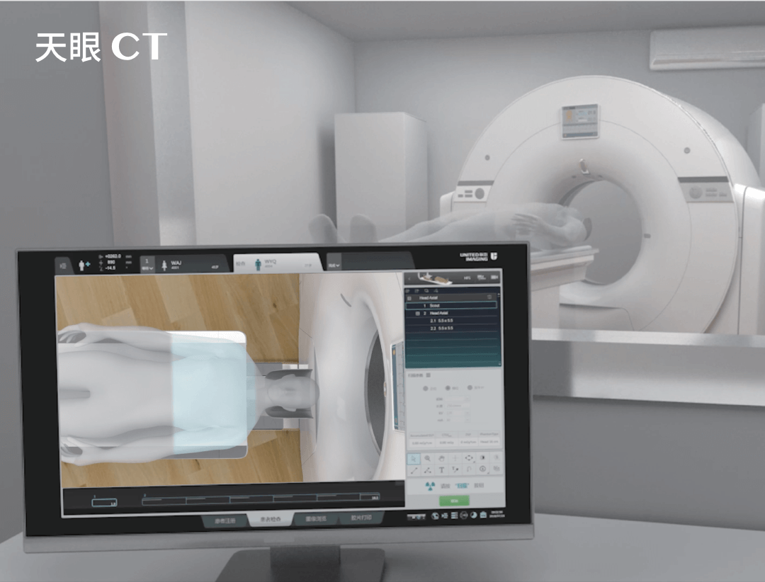
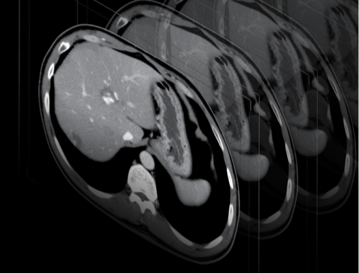
Computed tomography – precise imaging of internal structures
Computed tomography (CT), which combines X-rays with computer algorithms, produces detailed cross-sectional (2D and 3D) images of the part of the body being scanned. This method is extremely useful for diagnosing pathologies of the central nervous system, lungs, abdominal cavity and pelvic organs. A CT scan provides a quick and accurate assessment of a patient’s condition, but involves a higher dose of ionising radiation than an X-ray.
MRI – advanced imaging without radiation
Magnetic resonance imaging (MRI) is one of the most advanced diagnostic imaging techniques, using strong magnetic fields and radio waves to produce detailed images of the body’s internal structures. MRI is particularly useful for diagnosing soft tissues, joints, the vascular system and the central nervous system.
An MRI scan is safe and does not involve ionising radiation, but can be stressful for claustrophobic patients. MRI requires the patient to lie still in the machine during the scan, which can take between 15 and 90 minutes. Despite the considerable sound pressure level generated by the machine, the examination is painless and safe for the patient.
Nuclear medicine – specialised functional imaging
Nuclear medicine (PET) is a specialised branch of diagnostic imaging in which small amounts of radioactive isotopes are administered to a patient. Detectors placed outside the body record the gamma rays emitted by these isotopes, allowing the function of organs and tissues to be assessed. These scans are particularly useful for diagnosing the function of organs such as the kidneys and thyroid gland, as well as locating areas of inflammation and tumours.
Although advanced, nuclear medicine is not widely used as a standard diagnostic modality due to its high cost and limited availability. However, it is a valuable adjunct to other imaging modalities in special diagnostic cases.
Importance of imaging in medicine
Diagnostic imaging has played a key role in the development of modern medicine, providing doctors with an accurate understanding of a patient’s condition. It offers unparalleled capabilities in diagnosing a wide range of conditions from simple bone fractures to complex cancers and neurological diseases. Imaging allows doctors to plan more effective treatment strategies, monitor treatment progress and provide preventive healthcare.
Imaging tests are advanced and versatile, but unfortunately access to them is still limited in many parts of the world. Providing equal access to these key diagnostic tools, which are essential to the delivery of quality healthcare worldwide, remains a challenge, and it is the mission and goal of United Imaging Healthcare to address this challenge.
*ATTENTION! The information contained in this article is for informational purposes and is not a substitute for professional medical advice. Each case should be evaluated individually by a doctor. Consult with him or her before making any health decisions.