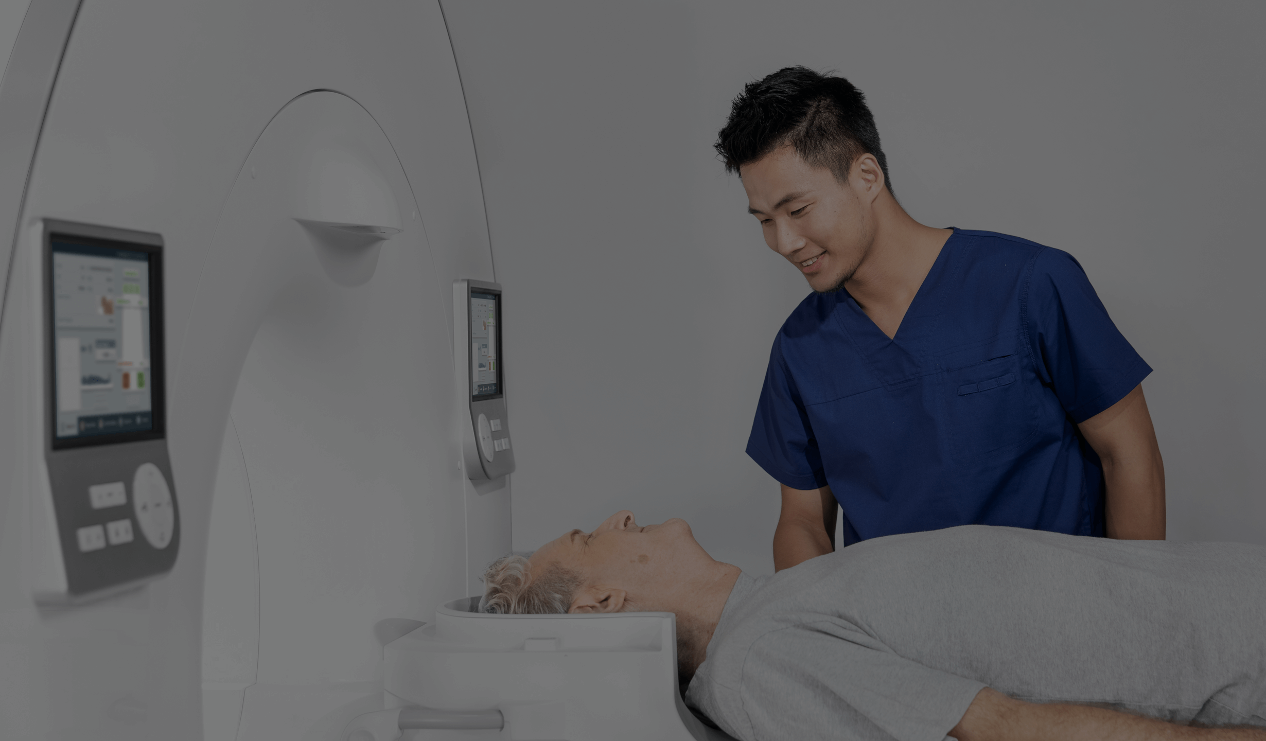
Head MRI scan
This painless and non-invasive method provides vital information that can be key to the diagnosis and treatment of many conditions.
What does a head MRI scan look like?
Head MRI uses the physical properties of hydrogen atoms in the body. When placed in a strong magnetic field, the protons in hydrogen atoms align in parallel and are subsequently excited by radio waves. When the protons return to their original state, they emit signals that are recorded and transformed into precise images by modern computer software.
What structures can be imaged by head MRI?
MRI of the head allows for detailed imaging of the brain, brain tissue, nervous system and blood vessels as well as orbital and pituitary structures, which makes it an indispensable tool in diagnosing neurodegenerative diseases, inflammation, cancer and head injuries.
SEE ALSO:
Types of head MRI
In addition to whole-brain MRI, depending in the structures that are analysed in detail, certain dedicated types of head MRI can be distinguished:
- Pituitary MR – a specialised MRI examination that focuses on evaluating the pituitary gland, which a small but very important structure at the base of the brain. This examination is used to diagnose pituitary gland tumours, cysts and other endocrine disorders.
- MR of the orbit/optic nerve – orbital MRI is an advanced diagnostic tool for precise assessment of the health of the eyes and surrounding structures, allowing the detection of various pathologies and monitoring of the treatments undertaken. It can also be focused on the optic nerve to diagnose its inflammation, tumours and other damage to the optic nerve.
- Craniofacial MRI – an MRI examination focusing on the craniofacial bones, temporomandibular joints, paranasal sinuses and soft tissues of the face. It is used to diagnose sinus disorders, jaw abnormalities, craniofacial tumours and trauma.
- MR of the inner ear and vagus structures – MRI of the inner ear and vagus is used to diagnose the causes of hearing loss, dizziness and other balance disorders. It can detect tumours such as auditory neuroma and other abnormalities of the inner ear and semicircular canals.
- MR angiography of the head – a specialised form of MRI focused on blood vessels in the head, including arteries and veins. It enables diagnosis of aneurysms, vascular malformations and cerebral venous thrombosis.
- Functional magnetic resonance imaging of the head (fMRI) – allows brain activity to be imaged by detecting changes in blood flow in response to neuronal activation. The examination is used to map brain function before surgery and in functional brain studies, including cognitive neurology.
- MR diffusion (DWI) and diffusion tensor imaging (DTI) – this examination enables assessment of the movement of water molecules in brain tissue, which is useful in the rapid diagnosis of ischaemic strokes. Diffusion tensor imaging (DTI) is an advanced form of DWI that can image nerve fibre pathways in the brain; this examination is useful in diagnosing neurodegenerative diseases and assessing damage following head trauma.
- Head perfusion MRI – this examination analyses blood flow in the brain, which is crucial in diagnosing and evaluating ischaemic strokes and brain tumours. This scan makes it possible to determine which areas of the brain are not receiving enough blood.
Each of the head MRI scan types listed above offers a unique perspective on the structures being examined and can be tailored to the patient’s specific diagnostic needs. It should be noted that state-of-the-art MRI scanners from United Imaging Healthcare enable the most advanced head MRI examinations to be conducted.
SEE ALSO: ABDOMINAL MRI SCAN
What can an MRI of the head detect?
A head MRI scan is effective in detecting the following conditions:
- brain disorders and malformations;
- cranial blood circulation disorders;
- vascular diseases;
- brain tumours;
- orbital diseases;
- meningitis;
- pituitary diseases;
- neurodegenerative diseases.
Head MRI scan – indications
When is an MRI of the head performed? The most common indications for this examination are recurrent headaches and migraines, balance and coordination disorders, vision problems and head injuries. A head MRI scan is also invaluable in the diagnosis of proliferative lesions, including brain tumours or neurodegenerative diseases such as multiple sclerosis, Parkinson’s or Alzheimer’s disease, and in monitoring treatment effects.
A decision to conduct a head MRI is usually preceded by a medical consultation, during which the doctor evaluates the patient’s symptoms and medical history to determine whether magnetic resonance is the right imaging modality in this case.
*ATTENTION! The information contained in this article is for informational purposes and is not a substitute for professional medical advice. Each case should be evaluated individually by a doctor. Consult with him or her before making any health decisions.
SEE ALSO: CRANIOFACIAL COMPUTED TOMOGRAPHY


