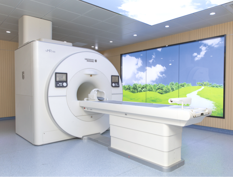
MRI of the hip
Because of its anatomical structure and the daily stresses it is subjected to, the hip joint is susceptible to a variety of injuries and diseases. One of the most effective ways to diagnose conditions of this joint is MRI, which is completely non-invasive and painless. It is perfect both as a stand-alone diagnostic method and as an adjunct to other diagnostic procedures. It uses a magnet, radio waves and an advanced computer to produce extremely accurate and detailed images of the inside of the hip joint.
What is the hip joint?
The hip joint is made up of the femoral head and the acetabulum. Its structure includes strong ligaments and a joint capsule, as well as many muscles that surround it. The hip joint is crucial for proper movement, including walking and running.
What is an MRI of the hip?
An MRI of the hip is a very valuable diagnostic tool that allows accurate imaging of this complex anatomical structure. A non-invasive test, MRI provides high-resolution images to help doctors diagnose various diseases and injuries and develop effective treatment plans.
What conditions can an MRI of the hip detect?
There are many indications for an MRI of the hip joint, as this examination provides valuable information about the condition of this complex anatomical structure. The main indications include:
- Hip pain – people with hip pain can have a variety of problems including arthritis, injury or degenerative changes. An MRI scan can help pinpoint the exact source of the pain.
- Reduced joint mobility – problems with the range of motion of the hip joint can be caused by a number of conditions, such as osteoarthritis or soft tissue damage. An MRI scan can help assess the extent of the damage and plan appropriate treatment.
- Morning stiffness in the hip – a feeling of stiffness, especially after a prolonged period of immobility, may indicate inflammation or other pathology in the joint. An MRI scan allows assessment of the soft tissues, which can help make a diagnosis.
- Swelling in the hip joint – swelling can be the result of injury, inflammation or degenerative disease. An MRI scan allows detailed imaging of the tissues, making it easier to identify the cause of the swelling.
- Bruising and haematomas around the hip – mechanical injuries, such as falls, can cause bruising and haematomas that may require further diagnosis. MRI helps to assess the damage and condition of the tissues around the hip joint.
What are the indications for an MRI of the hip?
MRI imaging of the hip joint is usually recommended for a wide range of conditions and symptoms that require accurate diagnosis and assessment of the hip joint. The main indications for this scan include:
- chronic pain that does not go away and may be the result of trauma, inflammation, degenerative disease or other pathology of the hip joint;
- various hip injuries, where detailed images obtained during the examination are important in determining the extent and nature of the damage, which is crucial for appropriate treatment;
- an MRI scan may be ordered both for diagnostic purposes and prior to surgery, as well as to monitor the progress of treatment for degenerative diseases;
- other conditions such as necrosis of the femoral head, tumours or acetabular-femoral conflict, which may also require detailed diagnosis.
MRI of the hip joint – contraindications
There are several absolute contraindications to MRI of the hip joint. These contraindications include patients with pacemakers, neurostimulators, orthodontic braces and orthopaedic screws, dental implants and certain types of fillings, contraceptive IUDs, shrapnel, bullet fragments, or even the presence of iron filings in the body if the patient is a locksmith or turner. For pregnant patients, the doctor may postpone an MRI scan, especially in the first trimester of pregnancy. The final decision is made by the doctor, who should consider all the pros and cons of having the scan.
How to prepare for a hip MRI?
Magnetic resonance imaging does not require any special preparation from the patient if it is done without contrast. In this case, the referring physician will order additional tests, such as blood creatinine levels to check kidney function. It is also advisable for the patient to arrive approximately 15 minutes before the scheduled appointment. The patient should wear comfortable, loose clothing without any metal parts, and remove jewellery and similar items. The mobile phone should be switched off. All personal belongings can be left in the designated area. In the case of joint MRI, it is not necessary to fast before the examination. It is a good idea to bring the patient’s existing medical records.
MRI of the hip joint – what does the examination look like?
A patient undergoing an MRI section of the hip will be under the constant care of medical staff. If necessary, a contrast agent is given to help visualise the structures being scanned. The patient is subsequently placed on a table that slides inside the scanner. To improve patient comfort, facilities around the world are purchasing state-of-the-art scanners, such as the uMR 570 from United Imaging Healthcare, which enables gantry diameter to be customised to individual needs. The scan takes between 15 and 45 minutes, during which time the patient must remain still. The scanner includes a microphone so the patient can alert the staff to any problems.
*IMPORTANT! The information contained in this article is for informational purposes only and is not a substitute for professional medical advice. Each case should be evaluated individually by a doctor. Consult with your doctor before making any health decisions.
