Pelvic CT scan – what is it, who is it for and how should I prepare?
A pelvic computed tomography scan is an advanced imaging test that uses X-rays and computer data processing to enable a detailed assessment of pelvic structures including bones, joints, muscles, ligaments as well as internal organs such as the bladder, uterus, ovaries and prostate. It is a key diagnostic tool that can detect damage and various pathological conditions potentially affecting the function of the pelvis and surrounding structures.
How does a CT scan work?
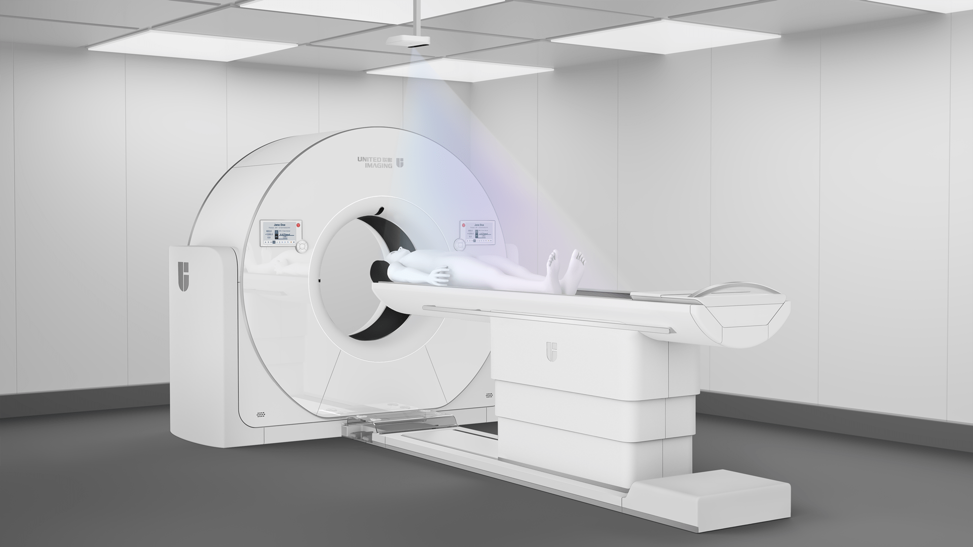
Computed tomography uses X-rays to produce detailed images of the body’s internal structures. During the examination, the patient lies on the moving table, which moves through the ring-shaped gantry of the CT scanner. Radiation that passes through the patient’s body is picked up by moving detectors. The computer processes these data to create cross-sectional images, which constitute scans of various pelvic structures. Compared to traditional X-rays, computed tomography provides more detailed information, allowing assessment of both soft tissues and bones.
Indications to the examination
Pelvic CT scans are indicated in a variety of symptoms and conditions that may require detailed diagnosis. Indications for pelvic CT scans include:
- pain and discomfort in the pelvic area;
- diagnosis and monitoring of tumours of the pelvic organs, such as prostate cancer, ovarian cancer, and uterine fibroids;
- identification of inflammatory conditions, such as pelvic organ inflammation, abscesses or infections;
- assessment of traumatic injuries to pelvic bones, joints and soft tissues;
- diagnosis of endometriosis, ovarian cysts, prostate problems and other internal organ disorders.
Additionally, pelvic CT is invaluable in planning surgical interventions, as it provides high-resolution images that enable lesions to be accurately located and the most effective treatment to be planned.
Contraindications to a pelvic CT scan:
Pelvic CT is a safe examination, but there are certain situations in which this imaging modality requires additional consultation or it cannot be used. These contraindications include:
- the period between the 3rd and 15th weeks of pregnancy – due to the use of X-rays, the examination should be avoided in pregnant women unless absolutely necessary;
- severe renal disease – especially where contrast agent is administered, which can impair kidney function;
- acute circulatory failure;
- hypertension;
- insulin-dependent (type 1) diabetes.
The examination is also not performed in children under the age of 2 and in elderly people over the age of 60. Not all the above contraindications mean that CT diagnostics is completely ruled out, and therefore the referring physician and the medical staff performing the examination should be aware of the patient’s medical conditions.
Who can order this examination?
A CT scan can be ordered by any doctor. In the case of examinations reimbursed by the National Health Fund, there is another restriction, since such reimbursement is only possible if the referral for the examination is issued by a specialist.
How to prepare for a CT scan
On the day of the examination, the patient should wear comfortable clothing without metal parts and remove all additional items such as jewellery or watches. If contrast agent is to be administered, the patient should be fasted for at least four to six hours before the examination.
Additionally, before an examination that involves the administration of a contrast agent, the patient may also be asked to undergo a creatinine level test to ensure that his or her kidneys are able to process the contrast agent.
When arriving for a pelvic CT scan, the patient should bring:
- the referral;
- his or her identity document;
- results of previous pelvic imaging studies, if applicable;
- creatinine test results, if ordered;
- other medical records pertaining to the area in question (e.g., hospital discharge summary reports, etc.).
Course of the examination
During the examination, it is important for the patient to remain motionless throughout the procedure, which usually takes a few to around a dozen minutes. If a contrast agent is required, it can be administered intravenously for better visualisation of pelvic structures.
After an examination that involves contrast administration, patients are advised to drink plenty of fluids, which helps the contrast agent to be removed from the body faster. Contrast administration may sometimes result in side effects such as nausea, rash, warmth, itchy skin or headache, although these are usually mild and transient reactions.
Pelvic CT results are usually available within a few days. The radiologist analyses the images and provides a report of the findings to the referring physician who will discuss the results with the patient and recommend further treatment, which may include medication, physiotherapy and sometimes surgical intervention.
Types of pelvic CT scans
A variety of computed tomography techniques are available for pelvic diagnosis purposes, which are tailored to the individual patient’s diagnostic needs. The basic examination enables the assessment of the overall structure and condition of the pelvic organs. Angio-CT, which is a specialised study of blood vessels, is an indispensable tool in the diagnosis of aneurysms or embolisms. 3D CT, on the other hand, is a perfect solution when the most detailed evaluation of anatomical pelvic structures is required, since it produces three-dimensional images, providing exceptional precision.
*ATTENTION! The information contained in this article is for informational purposes and is not a substitute for professional medical advice. Each case should be evaluated individually by a doctor. Consult with him or her before making any health decisions.