Chest MRI examination – what is it, who is it for and how should I prepare?
A chest MRI scan is a valuable modern medical diagnostic method, which enables precise and detailed imaging of the heart, lungs and other chest structures. Its safe and non-invasive nature, combined with the lack of exposure to ionising radiation, makes magnetic resonance imaging an effective diagnostic tool in a wide range of clinical cases, from diagnosing heart disease to evaluating trauma and cancer staging.
Indications for a chest MRI scan
MRI technology uses a powerful magnetic field and radio waves to produce detailed images that allow doctors to assess a patient’s health without undertaking invasive procedures. This study enables a detailed:
- diagnosis of congenital heart defects, cardiomyopathies and blood vessel problems;
- diagnosis of lung inflammations and other conditions, such as pneumonia and cancer;
- structural and functional evaluation of the aorta and other large blood vessels;
- diagnosis of thoracic masses that may indicate the presence of tumours;
- evaluation of chest condition after accidents and injuries, assessing damage to organs and bones.
Contraindications to the examination
Although chest MRI is a safe imaging technique that may be used in patients of all ages, there are certain situations where it may be contraindicated.
One contraindication is the presence of metallic implants in the patient’s body, such as joint replacements or stabilisers, which could affect the quality of MRI images as well as pose a potential risk to the patient due to the metal’s interaction with the strong magnetic field generated by the MRI scanner. In the case of metal implants, their type and material must be accurately determined before the examination.
Special attention should also be paid to patients with neurostimulators, implantable cardioverters-defibrillators or pacemakers. Although newer models of these devices are often designed to be MRI-compatible, older types may not be safe for use in a magnetic field. Each case requires individual consultation with a doctor and often also with the manufacturer of the device.
Metal vascular clips may pose a risk as well, especially if they are in close proximity to the area being examined. Before the examination, it must be ascertained that they are safe for the patient during MRI diagnosis.
Preparation for the examination
If a contrast agent is to be administered during the examination, a blood test for creatinine levels is crucial in order to assess the condition of the kidneys, which need to efficiently remove contrast from the body. For patients with any kidney disorders, the creatinine level test result should not be older than 7 days. For those with normal renal function, the maximum acceptable period between the test and the MRI scan is three weeks.
The patient should be fasting for several hours before the examination during which contrast agent is administered in order to minimise the risk of adverse reactions and provide better imaging conditions. If contrast administration is not necessary, no special diet is required.
On the day of the chest MRI examination, the patient should dress in clothing without any metal parts that could interfere with the images generated by the magnetic field of the MRI scanner. Any metal objects such as jewellery or glasses should also be removed before the examination.
Course of the examination
The patient should arrive at the medical facility a while before the appointed time to fill out the necessary documents and a questionnaire about his or her health and medical history. If a contrast agent is to be administered during the examination, an intravenous line (cannula) is inserted.
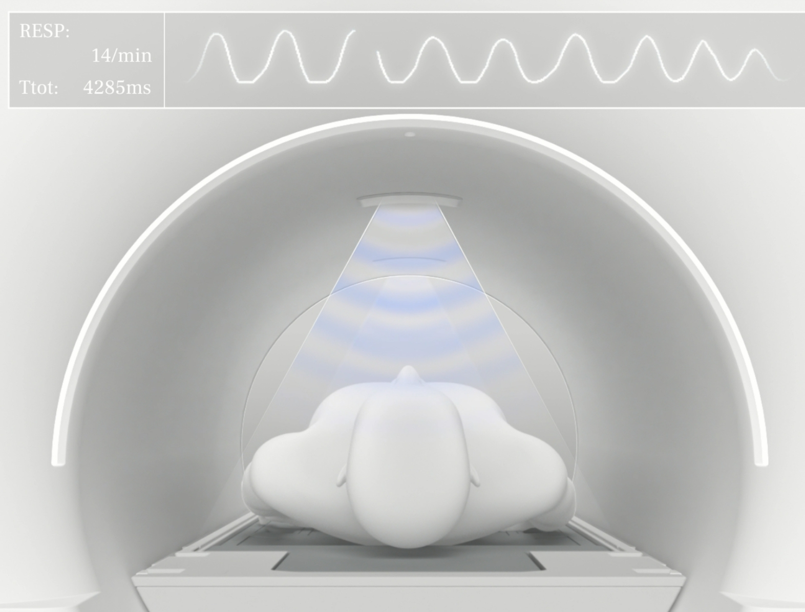
During the MRI scan, the patient lies on a moving examination table that is precisely positioned inside the MRI machine. It is important that the patient remain completely calm throughout the procedure and avoid any movements that could interfere with image quality. In some cases, especially when imaging vascular structures, the radiology technician may ask the patient to hold his or her breath for a few seconds to get clearer images.
The duration of a chest MRI scan can range from 30 to 60 minutes depending on the specific diagnostic indication.
Result interpretation
After the examination has been completed, the images are analysed by a radiologist who prepares a detailed report describing the results. Chest MRI result interpretation requires a great deal of experience and precision, since anatomical structures in this part of the body are complex and function dynamically. The radiologist evaluates the images for any abnormalities that may indicate the need for further diagnostic tests or medical interventions.
*ATTENTION! The information contained in this article is for informational purposes and is not a substitute for professional medical advice. Each case should be evaluated individually by a doctor. Consult with him or her before making any health decisions.


