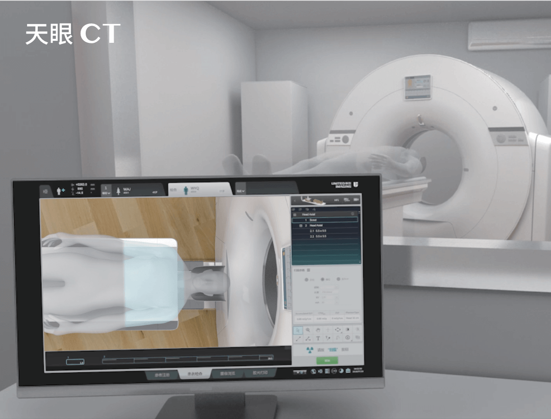
Spinal MRI scan
Magnetic resonance imaging of the spine is one of the most advanced imaging examinations, which allows a detailed assessment of spinal structures, including the vertebrae, intervertebral discs, spinal cord and surrounding soft tissues. Using a strong magnetic field and radio waves, MRI of the spine provides high-resolution images, which is crucial in the diagnosis of many conditions.
What does a spinal MRI scan look like?
A spinal MRI scan involves the excitation of hydrogen atoms in the body’s tissues using a strong magnetic field. Hydrogen protons emit signals, which are subsequently processed by a computer into images of cross-sections of the area examined. This enables doctors to obtain precise images of different parts of the spine – from the cervical through the thoracic to the lumbar section.
Structures imaged by a spinal MRI scan
As a multidimensional and multiplanar examination, MRI of the spine allows precise imaging of various structures and spaces, such as:
- intervertebral discs;
- the vertebrae and their shapes;
- the spinal cord and surrounding structures, including the nervous system;
- blood vessels;
- soft tissues around the spine, including muscles and ligaments.
SEE ALSO: ABDOMINAL MRI SCAN
Indications for a spinal MRI scan
An MRI scan of the spine is indicated in many situations. The most common indications for a spinal MRI scan are chronic back pain, symptoms indicative of pressure on nerve roots (radiculopathy), discopathies and spinal injuries, neurological disorders related to the spine (such as numbness or tingling in the limbs), suspected degenerative diseases or cancer, and monitoring of disease progression and treatment effectiveness.
What can a spinal MRI scan detect?
MR examination of the spine is a diagnostic method that can detect many different types of diseases and pathologies of the spine. These are usually:
- herniated and protruding intervertebral discs;
- inflammation;
- degenerative changes;
- spinal injuries;
- cancers;
- spinal cord and vascular diseases;
- demyelination characteristic of multiple sclerosis;
- congenital defects such as spina bifida.
What does a spinal MRI scan look like?
A spinal MRI scan usually takes 15 to 60 minutes. During the examination, the patient lies on a moving table that is moved inside the MRI scanner bore. It is important that the patient remain motionless so that the images obtained are as clear as possible. MRI of the spine can be performed with or without contrast, depending on the purpose of the examination. Many patients wonder whether it is necessary to undress for a spinal MR examination. If the clothes worn by the patient do not include metal parts, this is usually not necessary.
SEE ALSO: CT SCAN OF THE SPINE
Spinal MRI scan – types of specialised examinations
In the field of spinal MRI, several dedicated examinations are available that enable the structures of the spine and surrounding tissues to be accurately assessed. These advanced imaging techniques are extremely useful in diagnosing a wide range of spinal conditions from degenerative changes to trauma and cancer. These include primarily:
- MR diffusion-weighted imaging (DWI) of the spine – an examination that uses the molecular motion of water to generate images and is particularly useful in detecting early spinal lesions, such as inflammatory foci or tumours before these become visible on traditional MRI images. DWI can help distinguish between benign and malignant lesions and assess response to treatment.
- Functional MRI (fMRI) of the spine – although functional MRI (fMRI) is more commonly used to study the brain, under certain circumstances it can also be used to assess neural activity within the spinal cord, especially in studies of pain and its conduction. However, spinal fMRI is a more experimental technique and is not widely used in routine clinical practice.
- MR spectroscopy of the spine – this examination allows the chemical composition of tissue to be analysed, which can be helpful in distinguishing different types of tissue and lesions, including differentiation between tumours and non-tumour lesions. Similarly to spinal fMRI, MR spectroscopy is primarily a research tool.
- MR tractography of the spine – a method using diffusion tensor imaging (DTI), which allows visualisation and evaluation of nerve fibre pathways in the spinal cord. It is particularly useful in the diagnosis and evaluation of spinal cord injuries caused by both trauma and neurodegenerative diseases.
- MR myelography is a specialised examination that allows a detailed assessment of the subarachnoid space, spinal canal and spinal cord structures and nerves. It is particularly useful in the diagnosis of spinal stenosis (narrow spinal canal), disc herniations and other causes of pressure on the spinal cord and nerve roots.
Each of these examinations offers a unique perspective on spinal structures and can be selected depending on the patient’s specific diagnostic needs. It should be noted that state-of-the-art MRI scanners from UIH enable the most advanced spine MRI examinations to be conducted.
*ATTENTION! The information contained in this article is for informational purposes and is not a substitute for professional medical advice. Each case should be evaluated individually by a doctor. Consult with him or her before making any health decisions.
THIS MAY ALSO INTEREST YOU: HOW DO CONTRAST-ENHANCED SCANS WORK?


