Knee MRI scan
Magnetic resonance imaging of the knee is an advanced imaging technology that allows the structures of the knee joint to be analysed in detail. Owing to its high resolution and ability to accurately image soft tissue lesions, MRI is an indispensable tool in diagnosing sports injuries, degenerative diseases such as arthrosis as well as joint inflammation.
How does magnetic resonance imaging work?
MRI uses a strong magnetic field alongside radio waves. This combination makes it possible to obtain detailed images of the body’s internal structures. At the heart of the MRI scanner is a powerful magnet that is capable of generating a uniform, very strong magnetic field in the area to be examined.
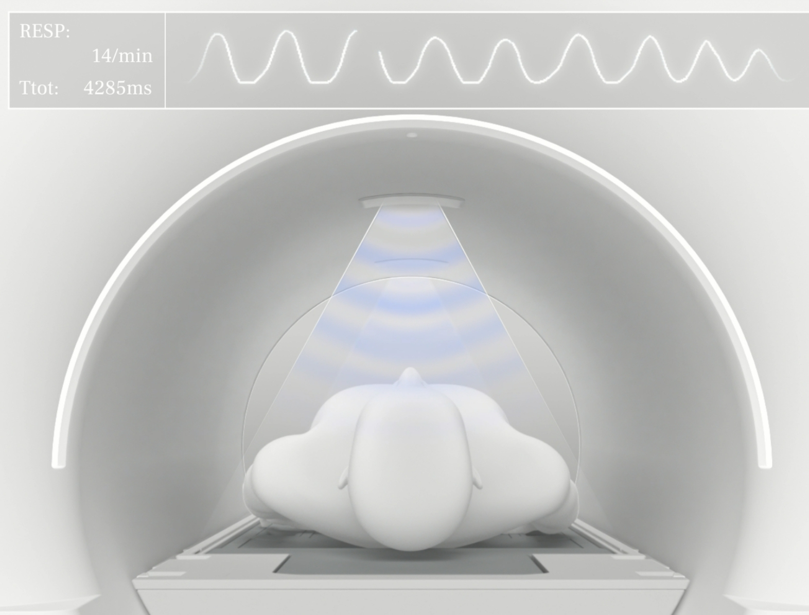
During the examination, the patient is placed inside a gantry that is an integral part of the MRI scanner. At this time, he or she is exposed to the aforementioned magnetic field.
The scanner emits short radio-frequency pulses using antennas installed in the MRI gantry, which are directed at the patient’s body. Hydrogen atoms in body tissues absorb these waves and emit magnetic responses in turn, which are then picked up by the scanner’s detectors.
This information is subsequently transferred to a computer, which processes it and converts it into detailed cross-sectional images of the examined area. This allows doctors to see very clear images of the tissue in question, which is crucial for accurate diagnosis and treatment planning.
Indications for knee MRI examination
An MRI examination of the knee joint is mainly performed to identify the causes of pain and restrictions in knee mobility, which are often early indications of problems with this joint. This examination is invaluable for diagnosing
- tendon injuries, ligament (ACL, PCL, MCL, LCL) injuries and meniscus tears. This is especially important for athletes, as accurate diagnosis is crucial for proper treatment and quick recovery;
- various joint inflammations. MRI images show any swollen tissue, synovial fluid and cartilage damage;
- when diseases such as osteoarthrosis are suspected. MRI can show the degree of cartilage degradation and the presence of free bodies in the joint space as well as making it possible to assess the condition of the bone and possible inflammation areas.
Contraindications to knee MRI examination
Although magnetic resonance imaging (MRI) of the knee is a safe and non-invasive procedure, there are some contraindications that should be considered before proceeding with the examination. These include:
- the presence in the patient’s body of metal implants, such as screws, plates or certain types of endoprostheses;
- the presence in the patient’s body of medical devices, such as pacemakers or cochlear implants, which may be damaged or interfere with the strong magnetic field generated by the MRI scanner;
- early stages of pregnancy;
- tattoos made with inks containing metal compounds.
In addition, patients suffering from claustrophobia (fear of enclosed spaces) may experience significant discomfort during the examination. In such cases, it is possible to use sedatives before the examination, but this must be consulted with the doctor.
In some cases, it is also necessary to administer a contrast agent, which is usually safe, but in patients with known renal insufficiency the use of contrast can lead to serious renal complications. In such cases, a detailed test of renal function is required before contrast administration.
Before undergoing an MRI of the knee, the patient should always carefully discuss all possible contraindications with the doctor or MRI technician. If there is any doubt about examination safety, alternative diagnostic methods should be considered.
Preparation for the examination
On the day of the examination, the patient should wear loose and comfortable clothing without metal parts. Jewellery, watches and other metal items may interfere with scanner operation.
If the administration of a contrast agent is planned, it is important to examine the patient’s renal condition before the examination, as some conditions may hamper the ability to excrete contrast from the body.
Course of the examination
The examination usually takes between 30 and 60 minutes depending on the purpose of the diagnosis. The patient is asked to lie on a table, usually in a supine position. The examined knee should be properly positioned, usually using special cushions or brackets that ensure stability and the correct angle. Positioning is key to obtaining the clearest images possible and it is customised depending on the individual examination. During the examination, the patient may hear loud, rhythmic knocking or clicking, which is the normal sound emitted by the scanner during operation. Communication with the radiology technician is possible via a microphone and speakers inside the MRI scanner. This allows the patient to report any problems or discomfort immediately, and enables the technician to inform the patient about the progress of the examination.
Following the MRI scan of the knee, the patient can return to normal activities unless otherwise instructed by the doctor. If the patient has been administered a contrast agent, it is advisable to drink plenty of fluids to assist its excretion from the body.
Examination results are usually available within a few days and will be forwarded to the attending physician who will discuss them with the patient, especially if any abnormalities have been identified that require further treatment or follow-up.
*ATTENTION! The information contained in this article is for informational purposes and is not a substitute for professional medical advice. Each case should be evaluated individually by a doctor. Consult with him or her before making any health decisions.


